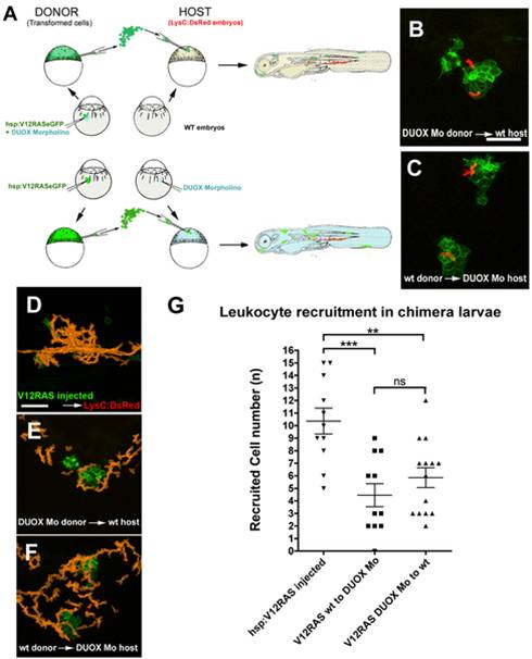
Fig. 7 Analysis of leukocyte recruitment in chimeric V12RAS-transformed cell-bearing larvae.
(A) Schematic diagram to illustrate how we generate chimeric V12RAS+ transformed cell-bearing Tg(LysC:DsRed) larvae, in which either the donor transformed cells or host tissues are morphant for DUOX. (B) A still image from Video S13B illustrating how LysC:DsRed+ cells are recruited to DUOX morphant V12RAS+ cells in WT host environment. (C) A still image from Video S13C showing LysC:DsRed+ cells recruited to WT V12RAS+ cells in a DUOX morphant host environment. (D–F) Cumulative LysC:DsRed+ cell footprints (orange) of representative movies shown in Video S13. (G) Graph to illustrate numbers of recruited LysC+ cells drawn towards V12RAS+ transformed cells in the two transplantation scenarios above compared with pTol2-hsp:V12RASeGFP injected into Tg(LysC:DsRed) control. **, p<0.01; ***, p<0.001; ns, not significant. Scale bars = 50 μm.

