
|
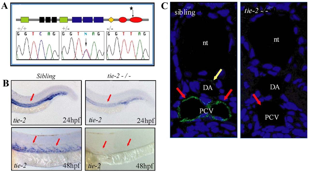
Fig. 1 A mutation in the kinase domain of tie-2 leads to loss of Tie-2 protein. (A) Schematic of the Tie-2 protein domains. Green, black and blue boxes represent the Ig-like, EGF-like and FN-III protein domains, respectively. The orange diamond represents the transmembrane region and the two red ovals represent the two kinase domains. Sequence chromatograms show the tie-2hu1667 allele to encode a stop codon within the second kinase domain. Sequence of the tie-2 hu1667 allele: CAG (Q982) is changed into a TAG (STOP) codon (mutation indicated by a star). (B) At 24 hpf, tie-2 mRNA expression levels are significantly reduced in tie-2 mutants compared with wild-type siblings (red arrows). tie-2 mRNA expression is absent at 48 hpf in mutants, whereas it is readily detectable in wild-type siblings (red arrows). (C) In wild-type fish, Tie-2 protein is expressed in the PCV at 48 hpf. Tie-2 protein expression is completely undetectable in tie-2 mutants. Yellow arrow shows Tie-2 protein expression in the dorsal aorta (DA); red arrows show Tie-2 protein expression in the PCV. nt, notochord.