
|
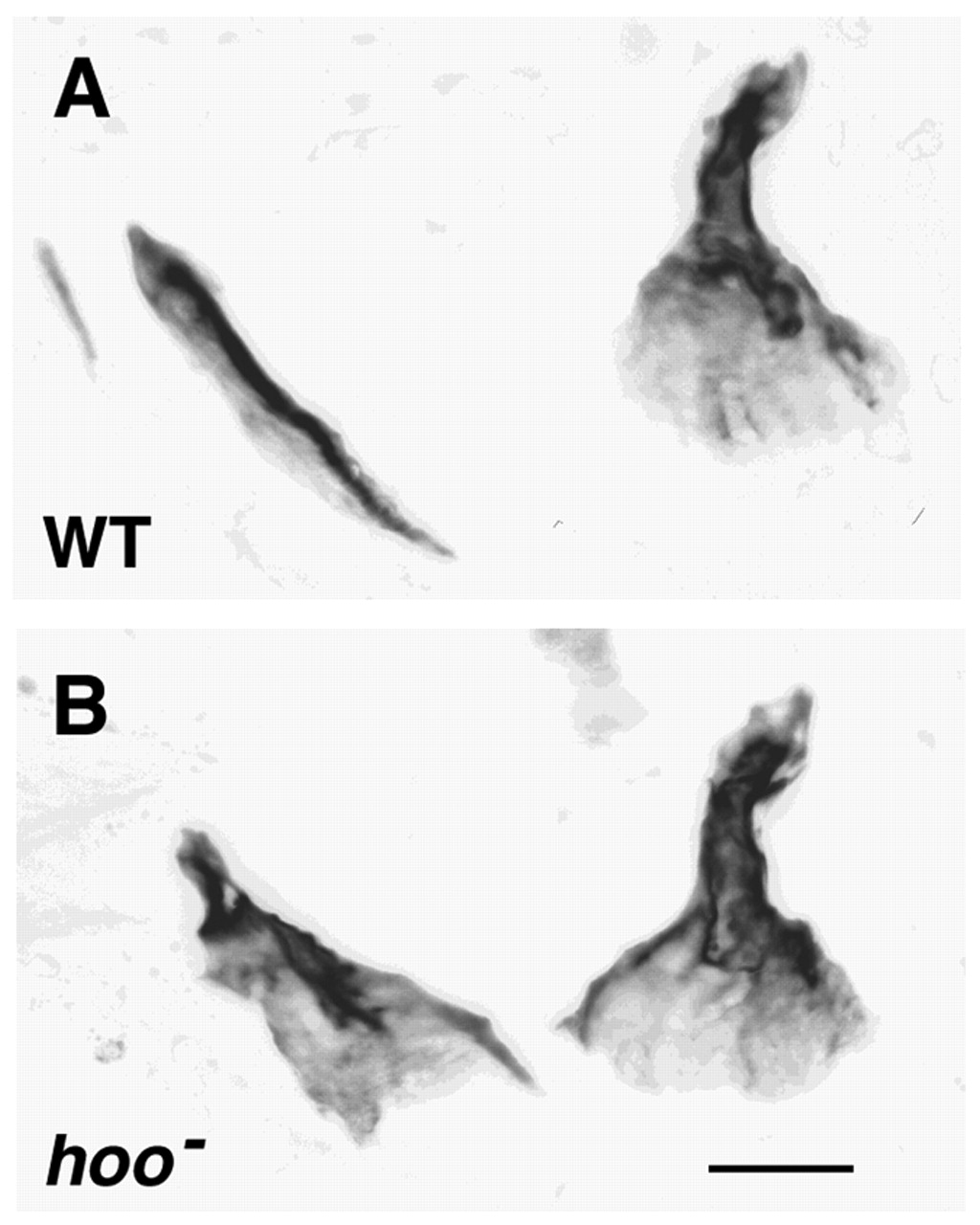
Fig. 9 Putative homeotic transformation of the dorsal branchiostegal ray towards the opercle in a hoover mutant. (A) A wild-type (WT) hoob631 sibling, showing the opercle and two branchiostegal rays, the more anterior one much smaller. (B) A hoo mutant. The opercle (right) is approximately normal in size and shape, but the bone at the usual position of the dorsal branchiostegal ray (left) has the form of an opercle. Its proximal end (upper) is sculptured into a distinctive joint region, rather than just ending bluntly as in A. Here DIC imaging (not shown; as in Fig. 4) reveals that the transformed branchiostegal ray makes a prominent joint with the underlying ceratohyal cartilage. Its distal region, normally blade-shaped, is expanded into an opercle-like fan. The other (more anterior and smaller) branchiostegal ray is missing. Scale bar: 50 μm.