
Fig. 3
- ID
- ZDB-IMAGE-110105-28
- Publication
- Tanaka et al., 2011 - Islet1 selectively promotes peripheral axon outgrowth in Rohon-Beard primary sensory neurons
- All Figures
- Figures for Tanaka et al., 2011
|
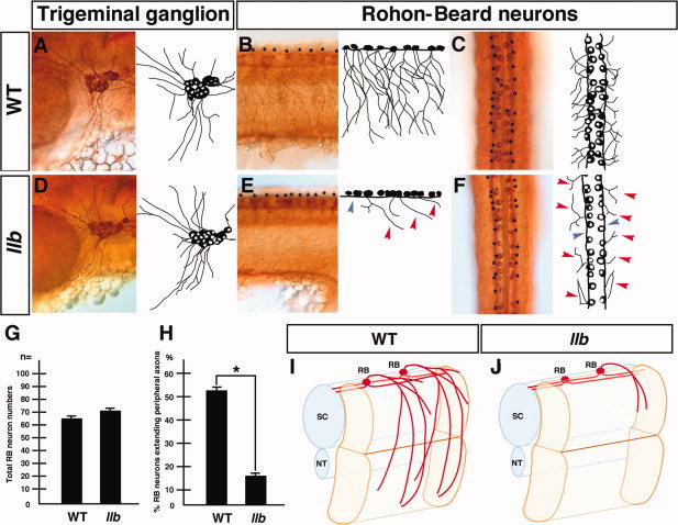
Fig. 3 llb mutation selectively affects the outgrowth of the peripheral axons of RB neurons. A–F: Primary sensory neurons of the wild-type and llb embryos at 28 hpf. Tg neurons (A, D) and RB neurons (B, C, E, F) were stained by anti-acetylated α-tubulin antibody. Lateral views with anterior at left (A, B, D, E); dorsal views with anterior at top (C, F). Red arrowheads, peripheral axons; blue arrowhead, central axons of RB neurons; asterisks, cell bodies of RB neurons. G, H: Quantitative data for the phenotypes of RB neurons in the wild-type and llb embryos. Data are mean ± SEM; n = 10; *P < 0.05, different from the wild-type embryos using 2-tailed paired Student′s t-test. I, J: Schematic views of the axial structure of the wild-type (I) and the llb embryos (J). NT, notochord; RB, Rohon-Beard neuron; SC, spinal cord.