
|
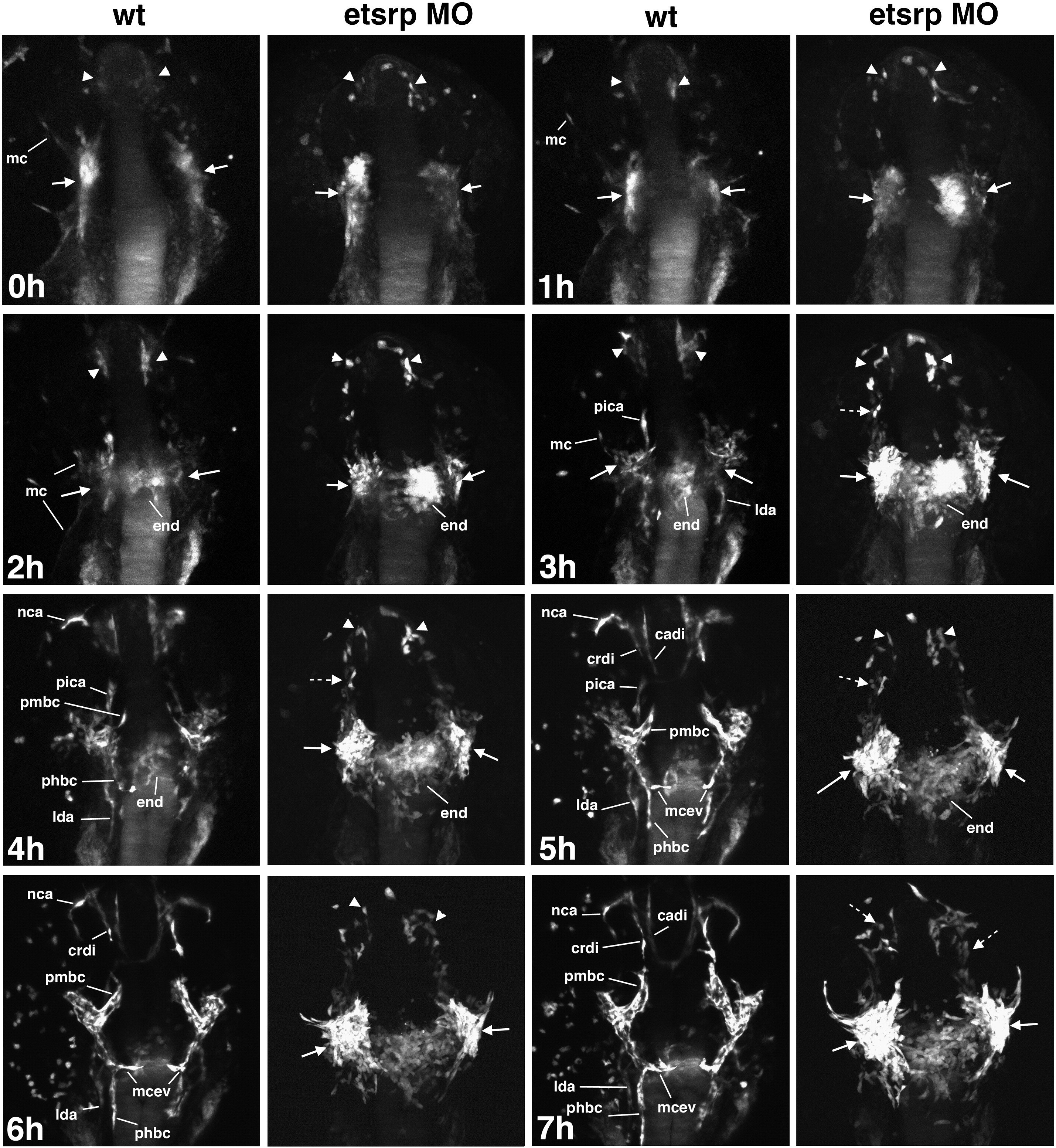
Fig. 9 Projection time-lapse images of cranial vessel formation in etsrp:GFP control (wt) and etsrp-MO injected embryos starting at approximately the 11-somite stage. Dorsal view of the anterior part of an embryo, anterior is to the top. 0–2 h: etsrp:GFP-expressing cells within ALPM coalesce into ROC (arrowheads) and MOC (arrows). Myeloid cells (mc) start to migrate out from MOC in wt embryos. In etsrp morphants, the initial ROC and MOC formation proceeds normally. No or very few migrating myeloid cells are present in etsrp morphants. 3–5 h: Angiogenic extensions that will give rise to cranial vessels including pica, phbc, lda and nca appear in wt embryos but not etsrp morphants. Significantly fewer myeloid cells are present in etsrp morphants than wild-type embryos (5 h panel). Note that multiple etsrp:GFP cells remain separated in etsrp morphants (dashed arrows) while all endothelial cell progenitors have migrated either to MOC or ROC in wt embryos. Migration of endocardial precursors (end) appears unaffected in etsrp morphants. 6–7 h: In wt embryos, ROC-derived crdi joins with MOC-derived pmbc. Angiogenic extensions of multiple other cranial vessels, including phbc, lda, mcev, cadi, and nca are apparent. In etsrp morphants, etsrp:GFP expression is bright and strong in the MOC area (arrows) where many endothelial cells originate but fail to migrate and only limited angiogenic extensions are apparent at these stages. Multiple endothelial cells in the ROC and in-between ROC and MOC remain separated and fail to coalesce into functional vessels (dashed arrows, 7 h).
Reprinted from Developmental Biology, 348(1), Proulx, K., Lu, A., and Sumanas, S., Cranial vasculature in zebrafish forms by angioblast cluster-derived angiogenesis, 34-46, Copyright (2010) with permission from Elsevier. Full text @ Dev. Biol.