
|
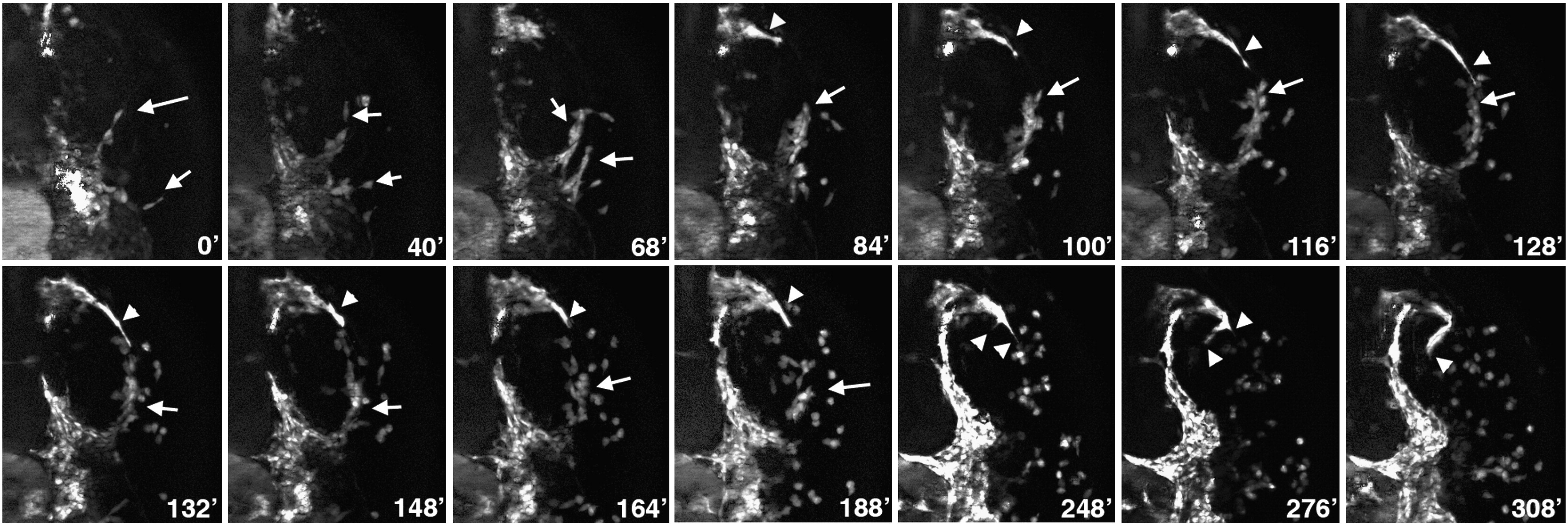
Fig. 7 Time-lapse images of myeloid cell migration. Dorsal view of the anterior right side of an embryo in the midbrain–forebrain area, anterior is up. Images start at the 14-somite stage; selected frames are shown. Note that the early myeloid precursors migrate in rows (arrows) and maintain cell–cell adhesion. A string of myeloid cell precursors migrates around the posterior-ventral side of the optic cup (arrow, 84–148 min). At the same time, endothelial precursors of nca vessel migrate at the anterior side of the optic cup (arrowheads, 84–188 min). During a short period of time, myeloid cells disperse and continue migration as individual cells (arrow, 164, 188 min). Nca vessel stalls, extends filopodia (arrowheads, 248 min) and then makes a 90° turn (276–308 min).
Reprinted from Developmental Biology, 348(1), Proulx, K., Lu, A., and Sumanas, S., Cranial vasculature in zebrafish forms by angioblast cluster-derived angiogenesis, 34-46, Copyright (2010) with permission from Elsevier. Full text @ Dev. Biol.