
|
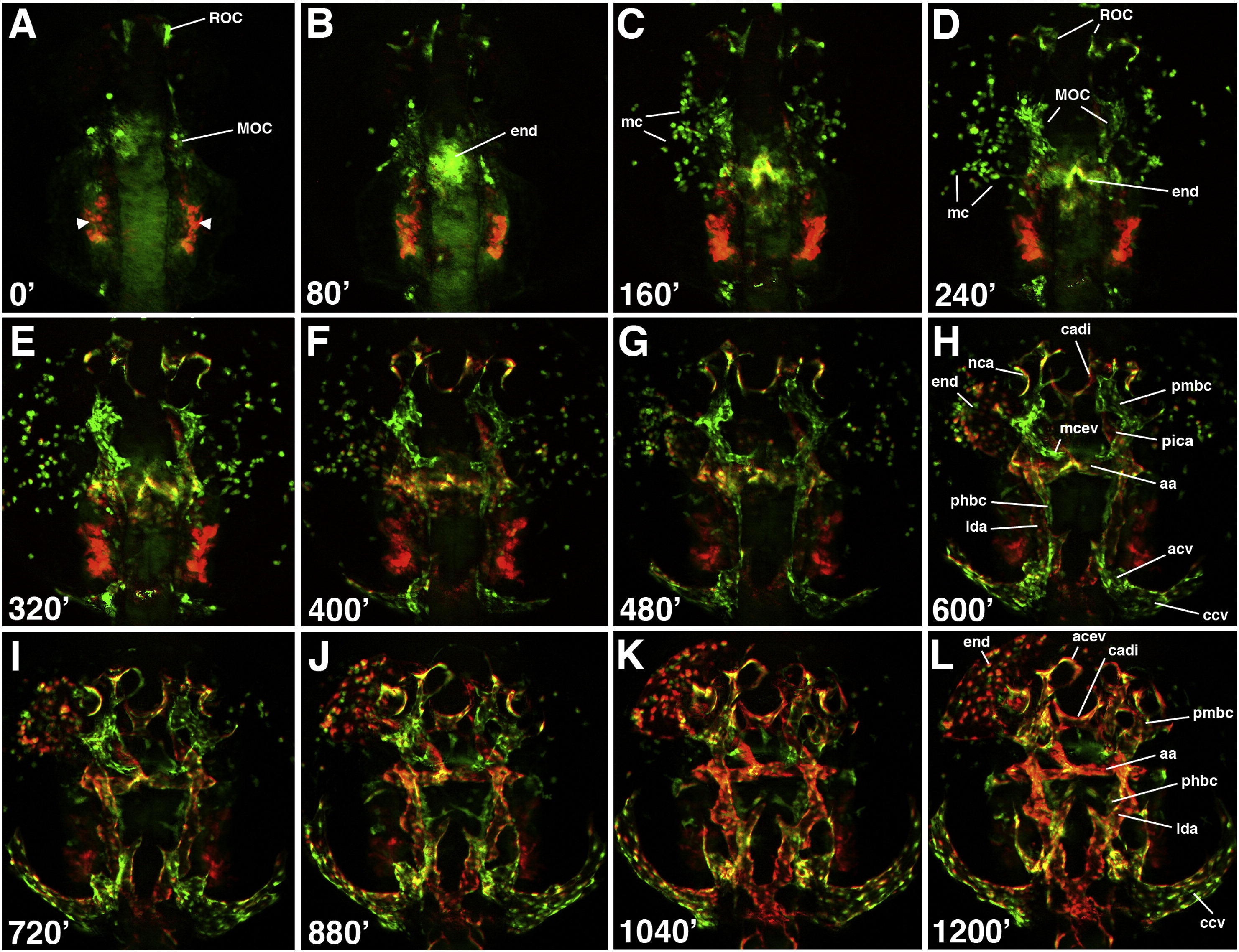
Fig. 6 Time-lapse images of cranial vessel formation in etsrp:GFP; kdrl:mCherry transgenic line from the 12-somite to approximately 32 hpf stages. Dorsal view of the anterior part of an embryo, anterior is to the top. (A–D) Cranial vessels originate from the rostral (ROC) and the midbrain organizing centers (MOC). Endocardial precursors (end) originate at the MOC and migrate to the midline by the 15-somite stage (B). Myeloid precursors (mc) migrate out of the MOC and disperse. There is very little kdrl:mCherry expression in cranial vessels during these stages. Most myeloid cells display only etsrp:GFP expression. Endocardial cells express both GFP and mCherry. Intense bilateral kdrl:mCherry expression posterior to the MOC is within pharyngeal endoderm. (E–H) Cranial vessels form angiogenic extensions from the ROC and the MOC. The rostral cranial vessels including acev and cadi originate at the ROC while mcev, phbc, pmbc and lda extend from the MOC. phbc and lda posterior extensions join with their counterparts that originate more posteriorly at acv/pcv and da and migrate anteriorly (G,H). Note that many venous vessels such as mcev, pmbc, and phbc display significantly stronger GFP expression and little or no mCherry expression while arterial vessels such as lda and aa display strong mCherry and weak GFP expression. The most rostral vessels such as acev and cadi express both GFP and mCherry. Endocardium which migrates to the left in (H) is positive for both reporter proteins while most myeloid cells express exclusively GFP. (I–L) Most cranial vessels lumenize and initiate circulation during these stages. The majority of cranial vessels now upregulate kdrl:mCherry expression and do not display significant differences in GFP and mCherry relative fluorescence levels.
Reprinted from Developmental Biology, 348(1), Proulx, K., Lu, A., and Sumanas, S., Cranial vasculature in zebrafish forms by angioblast cluster-derived angiogenesis, 34-46, Copyright (2010) with permission from Elsevier. Full text @ Dev. Biol.