
|
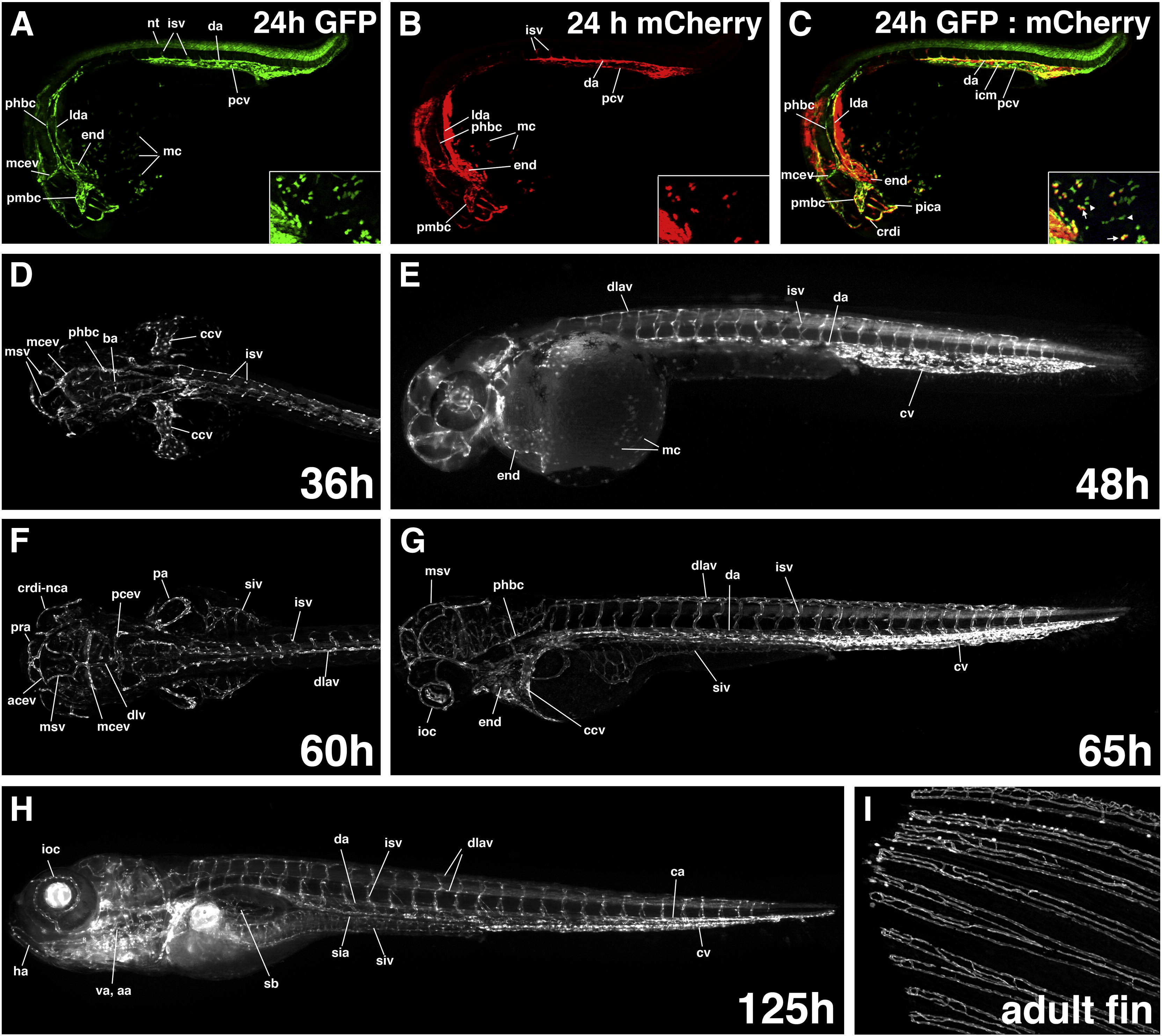
Fig. 2 Etsrp:GFP expression at 24 hpf and older embryos. (A–C,E,G,H) Lateral view, anterior is to the left; (D,F) dorsal view, anterior is to the left. (A–C) Comparison of etsrp:GFP (A) and kdrl:mCherry (B) expression at 24 hpf as observed in GFP (A), mCherry (B) channels and merged (C). Overlapping GFP and mCherry expression is observed in the dorsal aorta (da), posterior cardinal vein (pcv), intersegmental vessels (isv), cranial vessels that include lateral dorsal aorta (lda), primordial hindbrain channel (phbc), primordial midbrain channel (pmbc), middle cerebral vein (mcev), cranial division of internal carotid artery (crdi) and primitive internal carotid artery (pica) as well as the endocardium (end). There is apparent etsrp:GFP but not kdrl:mCherry expression in the intermediate cell mass region (icm) between da and pcv (C). Etsrp:GFP expression is present in multiple myeloid cells (mc) while flk1:mCherry is present only in some etsrp-expressing mcs (A–C, inset). GFP+mCherry- (arrowheads) and GFP+mCherry+ (arrows) mc populations are shown in the inset, C. Intense mCherry expression next to lda is in the pharyngeal endoderm. (D–H) Etsrp:GFP expression at 36 hpf (D, anterior part), 48 hpf (E), 60 hpf (F, anterior part), 65 hpf (G), 125 hpf (H). GFP fluorescence is apparent in all vascular endothelial cells that include da, pcv, isv, dorsal longitudinal anastomotic vessel (dlav), phbc, mcev, basilar artery (ba), mesencephalic vein (msv), common cardinal vein (ccv), anterior cerebral vein (acev), posterior cerebral vein (pcev), prosencephalic artery (pra), crdi-nasal ciliary artery (nca), dorsal longitudinal vein (dlv), inner optic circle (ioc), supraintestinal artery (sia), subintestinal vein (siv), hypobranchial artery (ha), ventral aorta (va), aortic arches (aa), pectoral artery (pa), swim bladder (sb), caudal artery (ca) and caudal vein (cv). GFP fluorescence is present also in the endocardium (end) and myeloid cells (mc). (I) Etsrp:GFP expression in the vasculature of adult caudal fin. Caudal fin edge is to the left.
Reprinted from Developmental Biology, 348(1), Proulx, K., Lu, A., and Sumanas, S., Cranial vasculature in zebrafish forms by angioblast cluster-derived angiogenesis, 34-46, Copyright (2010) with permission from Elsevier. Full text @ Dev. Biol.