
|
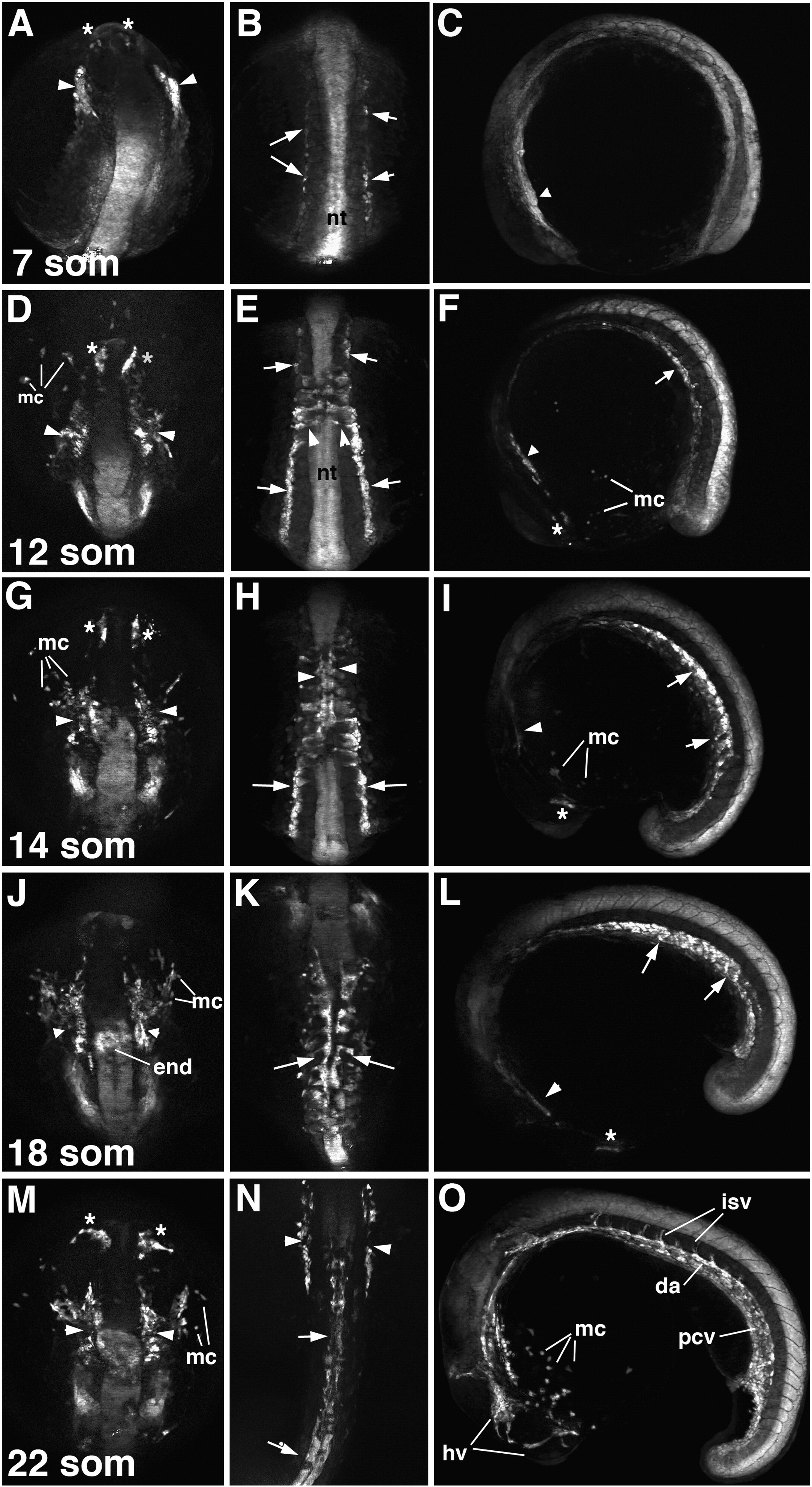
Fig. 1 Etsrp:GFP expression at the 7–22 somite stages. (A,D,G,J,M) Dorso-anterior view, anterior is up. (B,E,H,K,N) Dorsal view, anterior is up. (C,F,I,L,O) Lateral view, anterior is to the left. (A–C) 7-somite stage embryos. Note GFP expression in the endothelial and myeloid cell precursors within the rostral (ROC, asterisks, A) and the midbrain organizing centers (MOC, arrowheads, A,C) and in the angioblasts within posterior lateral plate mesoderm (PLPM, arrows, B). Neural tube (nt) displays non-specific GFP expression. (D–I) 12-somite (D–F) and 14-somite (G–I) stage embryos. Note GFP expression in the ROC (asterisks, D,F,G,I) and the MOC (arrowheads, D,F,G,I). Myeloid cells (mc) are migrating out from the ALPM. Bilaterally located GFP-expressing angioblasts within PLPM (arrows, E,F,H,I) migrate towards midline (arrowheads, E) where they aggregate into vascular cords to form axial vessels (arrowheads, H). (J–O) 18-somite (J–L) and 22-somite (M–O) stage embryos. GFP expression is apparent within endothelial and myeloid cell precursors in the ROC (asterisks, L,M) and the MOC (arrowheads, J,M) as well as in the endocardial precursors (end, J). Myeloid cells are migrating out of the ALPM. Angioblasts in the posterior part of an embryo aggregate into the vascular cords (arrows, K,L,N) that form the dorsal aorta (da) and the posterior cardinal vein (pcv). GFP fluorescence in intersegmental vessels (isv) is apparent. Intense GFP expression is observed at the rostral part of pcv (arrowheads, N). MOC and ROC-derived endothelial cells migrate to form head vessels (hv, O).
Reprinted from Developmental Biology, 348(1), Proulx, K., Lu, A., and Sumanas, S., Cranial vasculature in zebrafish forms by angioblast cluster-derived angiogenesis, 34-46, Copyright (2010) with permission from Elsevier. Full text @ Dev. Biol.