
|
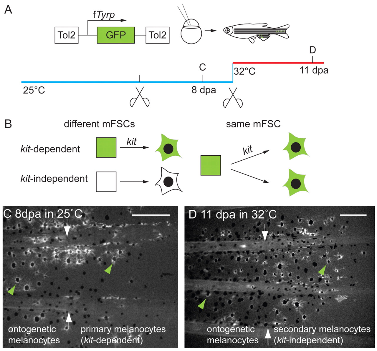
Fig. 7 kit-dependent and kit-independent regeneration melanocytes develop from the same mFSC. (A) kit-ts embryos were injected with the fTyrp>GFP lineage marker transposon and reared to adult stage at the permissive temperature. Following identification of fin ontogenetic and primary, kit-dependent regeneration melanocyte clones, fins were amputated again proximal to the original amputation plane and challenged to regenerate at the restrictive temperature for a further 11 days to reveal whether clones also contribute to kit-independent regeneration melanocytes. (B) How the kit-dependent and kit-independent regeneration melanocytes are related to each other: either they arise from different mFSCs (square), shown on the left; or they come from the same mFSC, shown on the right. (C,D) Fluorescence images of the same caudal fin clone regeneration (C) 8 dpa at 25°C and (D) 11 days after subsequent amputation and regeneration at 32°C. At the permissive and restrictive temperatures, GFP+ melanocytes (green arrowheads) regenerated distal to the GFP+ melanocytes in the stump. White arrows indicate amputation planes. Scale bars: 0.2 mm.