
Fig. 4
- ID
- ZDB-IMAGE-101124-5
- Genes
- Antibodies
- Publication
- Yin et al., 2010 - Hand2 Regulates Extracellular Matrix Remodeling Essential for Gut-Looping Morphogenesis in Zebrafish
- All Figures
- Figures for Yin et al., 2010
|
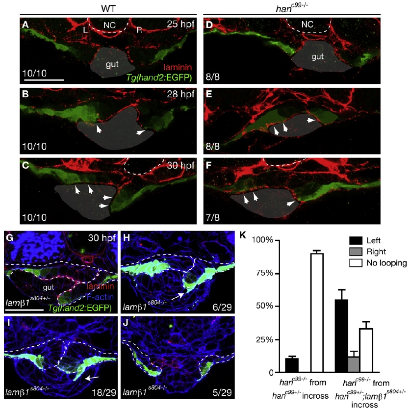
Fig. 4 hanc99 Mutants Exhibit Prolonged Deposition of Laminin at the LPM/Gut Boundary
(A–F) Time course analysis of Tg(hand2:EGFP) expression and laminin deposition during LPM migration. Ten wild-type (A–C) and eight hanc99 mutants (D–F) were fixed at the stages indicated and stained for laminin (red) and GFP (green). Dashed lines outline the notochord.
(A–C) In wild-type, laminin becomes diminished along the migratory path of Tg(hand2:EGFP)-expressing cells (arrows).
(D–F) In hanc99 mutants, laminin deposition persists along the entire LPM/gut boundary (arrows).
(G) The LPM in lamβ1s804 heterozygotes undergoes asymmetric migration (28/30).
(H–J) Out of 29 lamβ1s804 mutants examined, 6 showed incomplete asymmetric LPM migration (H), 18 had both the left and right LPM remain on top of the gut (I), and 5 had the LPM located lateral to the gut (J). Arrows point to the aberrant intrusion of LPM cells into the gut. Dashed lines outline the LPM.
(K) Percentages (mean ± SEM) of hanc99 mutants (n = 21) from hanc99+/- incrosses and hanc99 single mutants (n = 56) from hanc99+/-;lamβ1s804+/- incrosses showing gut-looping phenotypes according to foxa3 expression. Three independent crosses were examined.
All images are transverse sections, dorsal to the top. L, left; NC, notochord; R, right. The scale bars represent 40 μm. See also Figure S3.
Reprinted from Developmental Cell, 18(6), Yin, C., Kikuchi, K., Hochgreb, T., Poss, K.D., and Stainier, D.Y., Hand2 Regulates Extracellular Matrix Remodeling Essential for Gut-Looping Morphogenesis in Zebrafish, 973-984, Copyright (2010) with permission from Elsevier. Full text @ Dev. Cell