
Fig. 1
- ID
- ZDB-IMAGE-101124-2
- Genes
- Publication
- Yin et al., 2010 - Hand2 Regulates Extracellular Matrix Remodeling Essential for Gut-Looping Morphogenesis in Zebrafish
- All Figures
- Figures for Yin et al., 2010
|
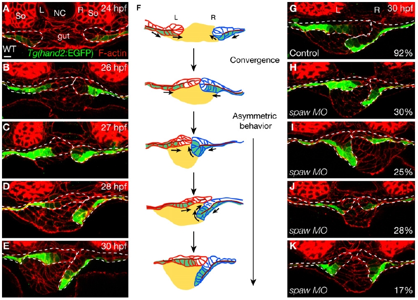
Fig. 1 Examination of Tg(hand2:EGFP) Embryos Reveals Novel Cell Rearrangements during LPM Migration
(A–E) Time course analysis of Tg(hand2:EGFP) expression in the LPM during gut looping. Ten Tg(hand2:EGFP) embryos were fixed every hour between 24 and 30 hpf, and stained for GFP (green) and phalloidin (red).
(F) Diagram of LPM migration. The gut is in yellow, the left LPM is in red, the right LPM is in blue, and the Tg(hand2:EGFP)-expressing cells are in green. The dark red line represents the apical side of the LPM epithelium. Arrows indicate the direction of migration of the Tg(hand2:EGFP)-expressing cells.
(G–K) Perturbing spaw function randomized the laterality of the rearrangements of the Tg(hand2:EGFP)-expressing cells. Compared to uninjected controls (G; n = 12), of 53 embryos injected with 10 ng of spaw MO, 30% showed the wild-type pattern (H); 25% showed the reverse pattern (I); 28% had both the left and right LPM migrate on top of the gut while the Tg(hand2:EGFP)-expressing cells on both sides remained ventral (J); and 17% had both the left and right LPM migrate ventrally and the Tg(hand2:EGFP)-expressing cells on both sides “rolled” dorsally (K).
All images are transverse sections, dorsal to the top. Dashed lines outline the LPM. L, left; NC, notochord; R, right; So, somite. The scale bar represents 10 μm.
Reprinted from Developmental Cell, 18(6), Yin, C., Kikuchi, K., Hochgreb, T., Poss, K.D., and Stainier, D.Y., Hand2 Regulates Extracellular Matrix Remodeling Essential for Gut-Looping Morphogenesis in Zebrafish, 973-984, Copyright (2010) with permission from Elsevier. Full text @ Dev. Cell