
|
Fig. 3
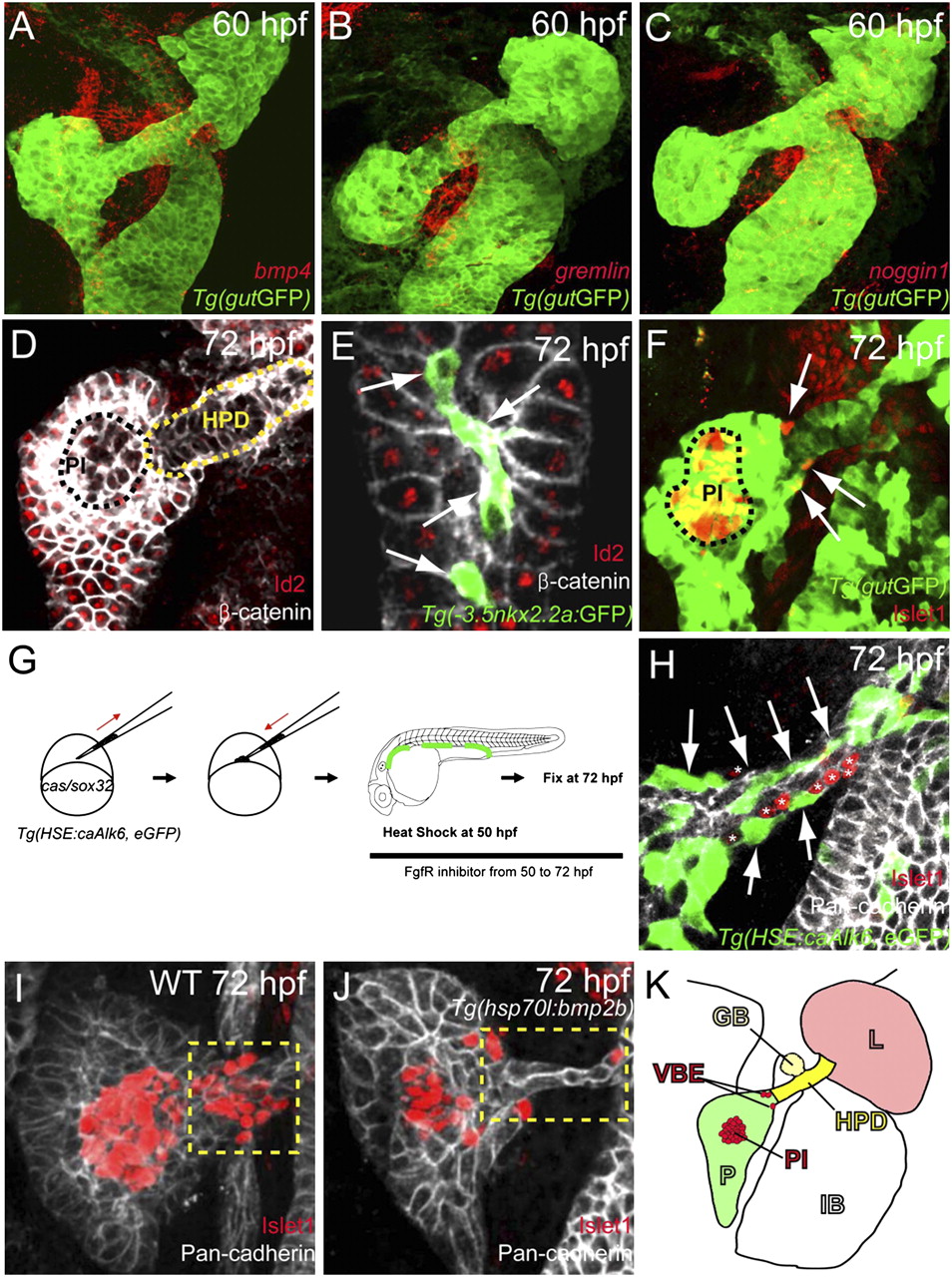
Activation of Bmp signaling cell-autonomously blocks the induction of ventral bud-derived endocrine cells. (A–C) Confocal images of Tg(gutGFP) (green) with mRNA expression (red) of bmp4 (A), gremlin1a (B), and noggin1 (C) at 60 hpf. (D and E) Confocal images of wild-type endoderm showing β-catenin (white) and Id2 (red) expression at 72 hpf. (D) Id2 expression is excluded from the hepatopancreatic duct cells (HPD; yellow dashed line) and appears to be downregulated or excluded from the pancreatic endocrine cells (PI, principal islet; black dashed line). (E) Id2 expression is also excluded from the intrapancreatic duct cells, labeled by Tg(-3.5nkx2.2a:GFP) expression (41) (green; arrows). (F) Confocal image of wild-type endoderm showing Tg(gutGFP) (green) and Islet1 (red) expression at 72 hpf. The ventral bud-derived endocrine cells (arrows) can be found at the junction between the pancreas and hepatopancreatic duct [the principal islet (PI) is outlined by black dashed line]. (G) Schematic diagram of the cell transplantation protocol. cas/sox32-overexpressing Tg(HSE:caAlk6, eGFP) donor cells were transplanted into wild-type hosts. Hosts were heat-shocked at 50 hpf and fixed at 72 hpf. After applying heat shock, hosts were treated with the Fgf receptor inhibitor SU5402, which induces ectopic Islet1-positive cells in the hepatopancreatic duct. (H) Single plane image of embryo stained for GFP (green), Pan-cadherin (white), and Islet1 (red) showing that Tg(HSE:caAlk6, eGFP)-expressing cells (arrows) fail to express Islet1 and display clear segregation from the ectopic Islet1-expressing cells (asterisks). (I and J) Effect of cell-nonautonomous activation of Bmp signaling by heat shock of Tg(hsp70l:bmp2b) at 50 hpf and subsequent treatment with SU5402 until fixation. Confocal projections of embryos stained for Pan-cadherin (white) and Islet1 (red) comparing control (I) and experimental (J) embryos. (I) Upon SU5402 treatment, ectopic Islet1-positive endocrine cells appeared in the hepatopancreatic duct (yellow dashed area), an effect that can be partially blocked by overexpression of Bmp2b (J). (K) Diagram of the endodermal organs at 72 hpf showing the liver (L; light red), pancreas (P; green), hepatopancreatic duct (HPD; yellow), gall bladder (GB; light yellow), and intestinal bulb (IB), as well as the ventral bud-derived endocrine cells (VBE; red) and principal islet (PI; red).