
Image
|
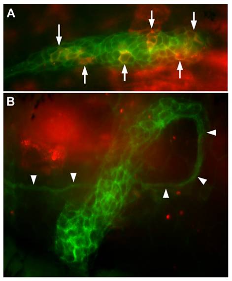
Figure Caption
Fig. S3 Transient expression driven by the 250-bp promoter in cldnb:gfp embryos (A) and in cldnb:gfp embryos injected with esr1 mRNA (B). A dozen cells are strongly fluorescent in A (arrows), and another 15 are more weakly fluorescent, as observed in half of the injected embryos. No cells are fluorescent in B, as observed in 85% of the injected embryos (see main text). Note the ectopic course of the primordium revealed by the nerve that follows primordium migration (arrowheads): the primordium first followed a normal course; then doubled dorsally and eventually U-turned to dive ventrally.
Acknowledgments
This image is the copyrighted work of the attributed author or publisher, and
ZFIN has permission only to display this image to its users.
Additional permissions should be obtained from the applicable author or publisher of the image.
Full text @ Proc. Natl. Acad. Sci. USA