
|
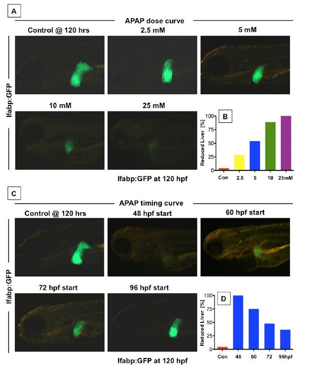
Fig. S4 APAP toxicity is dose- and time-dependent in zebrafish embryos. (A) lfabp:GFP transgenic embryos exposed to increasing doses of APAP at 72 hpf and assessed at 120 hpf showed progressively decreasing liver size. (B) Graphical representation of the fraction of zebrafish with diminished liver size at increasing APAP doses (n e 20 embryos for three replicates). (C) lfabp:GFP transgenic embryos were exposed to APAP, and in vivo fluorescence was assessed at 120 hpf. Embryos were exposed to 10 mM APAP at different time points during development. Decreasing time of exposure to APAP caused increasing liver fluorescence and liver size. (D) The fraction of zebrafish with decreased liver size declines with decreasing exposure time; n > 20 embryos/group with three repeats per treatment group.