
|
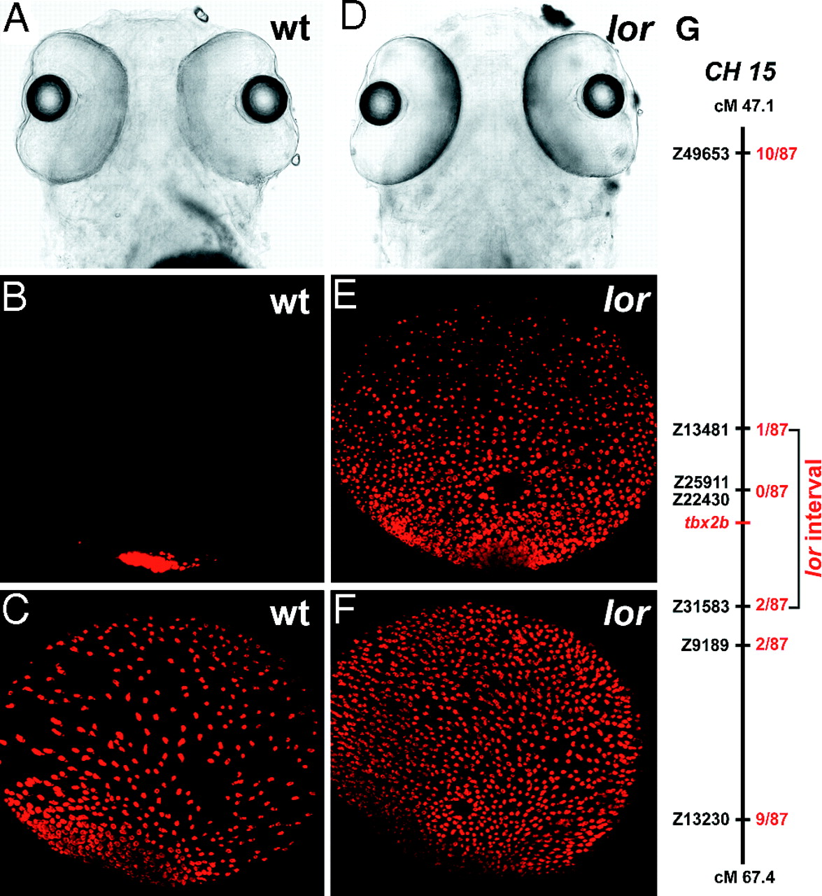
Fig. 1
lorp25bbtl mutants display increased labeling for rods. Ventral views of bright field images (A and D) and confocal immunofluorescent images of rod-specific labeling of eyes from WT (B and C) and a lorp25bbtl mutant larvae (E and F) (dorsal is up). At 3 dpf, WT larvae display rod labeling first in the ventral retina (B) followed at 5 dpf, by sporadic labeling of individual cells in the central and dorsal retina (C). At 3 dpf, lorp25bbtl mutant larvae display increased rod immunolabeling across the ventral and central retina (E) that is evenly distributed at 5 dpf (F). (G) Linkage analysis placed lorp25bbtl between SSLP markers Z31583 and Z13481 on the MGH panel. The number of recombinants in 87 mutant larvae is shown (red). The interval containing the lor mutation and the tbx2b locus is shown.