
|
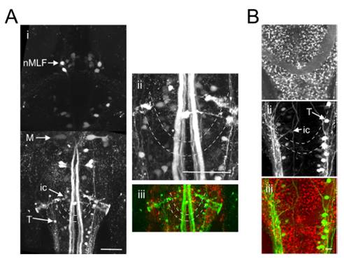
Fig. S7 The caudal reticular formation is located in the vicinity of the commissura infima Halleri. (A and B) Two Gal4s1101t; UAS:NpHR-mCherry animals in which a green dextran-conjugated dye was injected into the spinal cord to label reticulospinal cells. (Ai) Dorsal view, extending from the rostral spinal cord (Bottom) to the midbrain (Top). ic and T cells of the caudal reticular formation are located in close proximity to the commissura infima Halleri (dashed lines). M, mauthner cell, nMLF, nucleus of the medial longitudinal fasciculus. (ii) Magnified view of the region containing the ic and T cells. (iii) Merged channels image. (B) Red channel (i), green channel (ii) and merged channels (iii).