
|
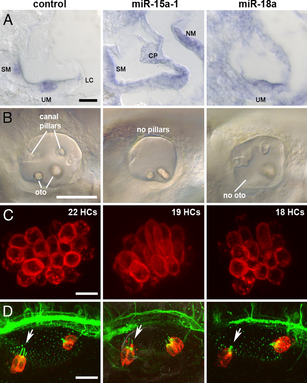
Fig. 4
miRNAs 15a and 18a are expressed in the zebrafish inner ear and miR-15a-1 or miR-18 morphant phenotypes include inner ear defects and reduced HC numbers. (A) ISH shows expression at 48 hpf: miR-183 served as a positive control (Left) for expression in HCs in the saccular macula (SM), utricular macula (UM), and lateral crista (LC). miR-15a was expressed in lateral line neuromasts (NM) and throughout the inner ear, including SCs and canal pillars (CP), and miR-18a expression was strong in the UM and adjacent cells. Dorsal is up, lateral is right. (B–D) Otocysts shown after treatment with MMO-miR-15a-1 or MMO-miR-18a (see Fig. S3). Injection with a standard MO that is not expected to target any known zebrafish RNA serves as a negative control (Left) to emphasize defects in canal pillars, otoliths (oto), and number of HCs in the other morphants. (B) Otocysts at 55 hpf. Anterior is left, dorsal is up. (C) UMs at 55 hpf. Confocal image stacks of HCs viewed en face from the ventral side. HC numbers are listed for each sample. Anterior is left, medial is up. (D) Otocysts at 30 hpf. Confocal image stacks show that morphants have differentiated the earliest HCs, called tether cells (red), at the anterior (arrow) and posterior poles. Primary cilia, labeled with acetylated tubulin, project into the lumen of the otocyst (green dots) but are substantially elongated for tether cells; axons also stain with this antibody. Anterior is left, dorsal is up. [Scale bars: (A), 20 μm; (B), 100 μm; (C), 10 μm; (D), 20 μm.]