
|
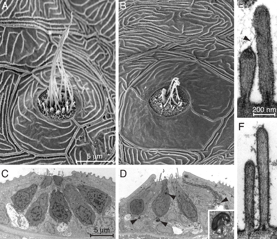
Fig. 5
Ultrastructural features of mutant and normal hair cells. (A) A scanning electron micrograph of a midtail neuromast from the posterior lateral line of a mutant at 6 dpf shows a dozen long kinocilia emerging from short clusters of stereocilia. (B) A neuromast from a similar position in an age-matched, homozygously mutant fish displays shrunken kinocilia and a diminished number of hair bundles. The semilunar periderm cells surrounding the neuromast are abnormally smooth. (C) A low-power transmission electron micrograph of a neuromast in a 6-dpf control larva shows parts of seven hair cells, some contacting large nerve terminals. (D) A mutant neuromast is characterized by several lysosomes (arrowheads) both within them and in supporting cells. A representative lysosome is enlarged fivefold in the inset. (E) A high-power micrograph from a control animal demonstrates a pair of sterocilia connected by a tip link (arrowhead) that terminates at its upper end in a prominent insertional plaque. (F) In a mutant larva of the same age, the stereocilia are narrow and display little basal tapering. Neither tip links nor insertional plaques are evident, and the shorter stereocilia lack dense material beneath their tips. The magnifications are identical for A and B, for C and D, and for E and F.