
Image
|
Figure Caption
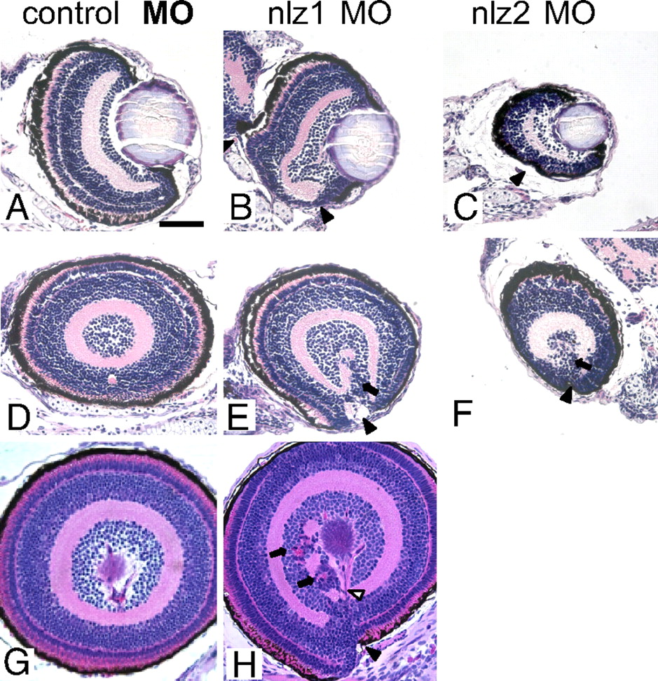
Fig. 3 Histopathology of nlz morphant fish. Coronal (A–C) and sagittal (D–H) planes through the zebrafish eye. Normal retinal lamination and a fused ventral fissure can be seen in control MO injected fish at 5 dpf (A and D) and at 6 dpf (G) (Top, dorsal; Bottom, ventral). Fissure closure defects can be seen in nlz1 morphant fish at 5 dpf (B and E) and 6 dpf (H) and nlz2 morphant fish at 5 dpf (C and F). The coloboma in morphant fish is accompanied by discontinuous RPE (black arrowhead), retinal dysplasia/rosettes (arrows), and abnormal vasculature (open arrowhead). H&E staining.
Figure Data
Acknowledgments
This image is the copyrighted work of the attributed author or publisher, and
ZFIN has permission only to display this image to its users.
Additional permissions should be obtained from the applicable author or publisher of the image.
Full text @ Proc. Natl. Acad. Sci. USA