
|
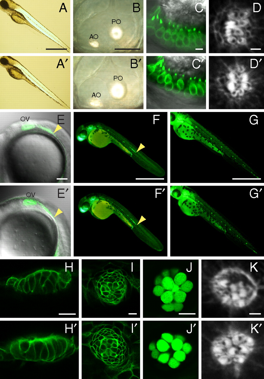
Fig. 2
Absence of gross morphological and developmental defects. In each panel, a WT larva (Upper) is contrasted with a mutant (Lower). (A, A′) Mutants display a normal body shape and length at 4 dpf. (B, B′) In a mutant, the semicircular canals develop normally and the anterior (AO) and posterior otoliths (PO) are normal. (C, C′) Labeling of hair cells by Tg(pou4f3:gap43-mGFP) (green) reveals no obvious differences in their number, shape, or localization in the anterior macula. (D, D′) In lateral-line neuromasts at 3 dpf, the hair bundles of mutants display normal planar cell polarity. The kinocilium of each hair bundle is marked by a black notch denoting the absence of actin staining by phalloidin. At 22 hpf (E, E′), 2 dpf (F, F′), and 3 dpf (G, G′), mutants display normal migration of the lateral-line primordium and formation of neuromasts, here labeled by Tg(cldnB:lynGFP) (green). OV, otic vesicle; arrowheads indicate the leading edges of the primordia. (H and I) Supporting cells labeled by Tg(cldnB:lynGFP) (green) are normally arranged in mutants in the anterior (H, H′) and posterior lateral lines (I, I′) of 5-dpf larvae. (J, J′) Fifty hours after treatment with Cu2+, a 7-dpf mutant larva shows normal regeneration of hair cells labeled by Et(krt4:GFP)sqet4 (green). (K, K′) Phalloidin staining shows that regenerated hair cells display planar cell polarity in a mutant. (Scale bars, 500 μm in A, F, and G; 50 μm in B; 5 μm in C; 2 μm in D and K; 100 μm in E; 10 μm in H–J.)