
|
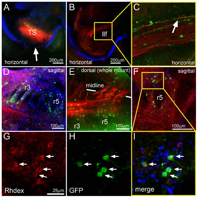
Fig. S4 Retrograde labeling of secondary octaval nucleus (SON) neurons. Adult brain cryostat sections (100 μm) of rhodamine dextran injected adult rh3/5:KalTA4 X 4xKaloop fish. (A) Rhodamine dextran (red) injection site in the central nucleus of the torus semicirculars (TS). The arrow demarcates the site where the tectum was removed. (B) Rhodamine dextran (red) positive axons innervate the TS coming from caudal projection nuclei and (C, enlarged area of B marked with yellow square) some are positive for GFP expression (white arrow). (D) Rhodamine dextran-positive axons are derived from hindbrain neurons, (E) they cross the midline at the level of r3 and r5 and (F) turn dorsally (G–I, enlarged area of F marked with yellow square) revealing their origin from GFP-expressing somata of rh3/5:KalTA4 X 4xKaloop labeled neurons. Abbr.: llf: lateral longitudinal fascicle, r: rhombomere, TS: torus semicircularis.