
|
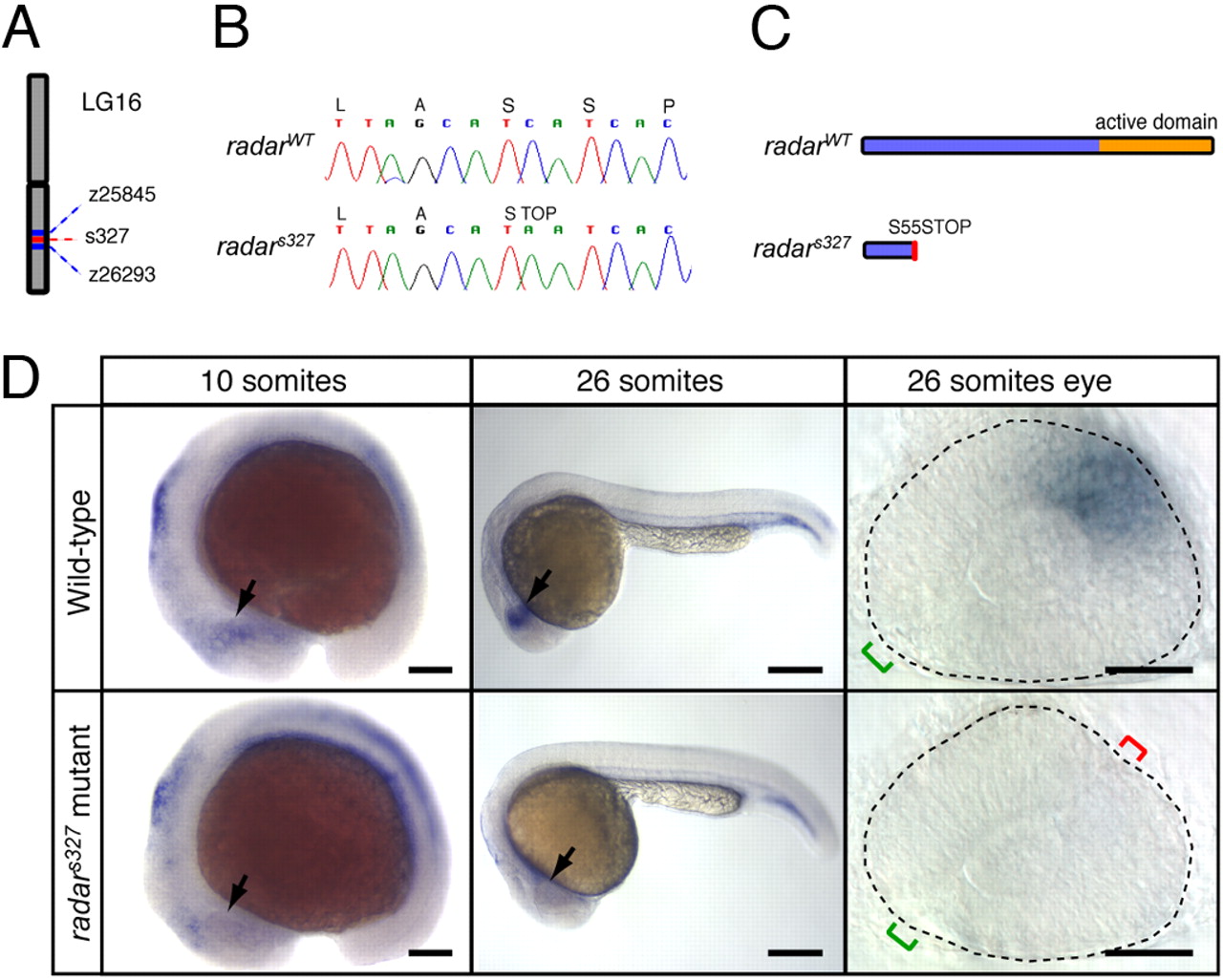
Fig. 2
Positional cloning and expression pattern of radar. (A) s327 maps to chromosome 16 between z25845 (2.3 cM) and z26293 (0.7 cM). (B) Sequencing of WT and s327 cDNA reveals a single C-to-A substitution in position 164 of the radar ORF, resulting in a premature stop codon. (C) Predicted translated peptides arising from radarWT and radars327. The mutation is predicted to result in a truncated protein, lacking the mature signaling domain. (D) Whole-mount in situ hybridization shows a restricted pattern of radar expression in WT embryos. radar mRNA is largely absent from the retina of radars327 mutants at all stages. In WT, expression is evident in the distal optic vesicle of WT embryos at 10 somites (arrow). At 26 somites, radar is expressed dorsally, opposite of the optic fissure (green bracket). Note ectopic fissures (red bracket) in radars327 mutants. (Scale bars: 150 μm for 10 somites, 250 μm for 26 somites, 50 μm for dissected 26 somite eyes.)