
Image
|
Figure Caption
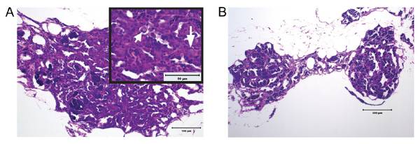
Fig. S5 Hyperplastic, dysplastic, and degenerative changes in H&E-stained sections of testes from adult brca2Q658X homozygous zebrafish. (A) Testicular tubules are lined by numerous germ cells that form multicellular layers (Inset, long arrow). Multifocally, dysplastic germ cells with large nuclei, vacuolated chromatin, and prominent nucleoli are seen (Inset, short arrow). (B) Testes from brca2Q658X homozygotes show segmental degeneration, with loss of spermatocytes in testicular tubules. (Scale bars: 100 μm in A; 200 μm in B; 50 μm in the Inset in A.)
Figure Data
Acknowledgments
This image is the copyrighted work of the attributed author or publisher, and
ZFIN has permission only to display this image to its users.
Additional permissions should be obtained from the applicable author or publisher of the image.
Full text @ Proc. Natl. Acad. Sci. USA