
Fig. 5
|
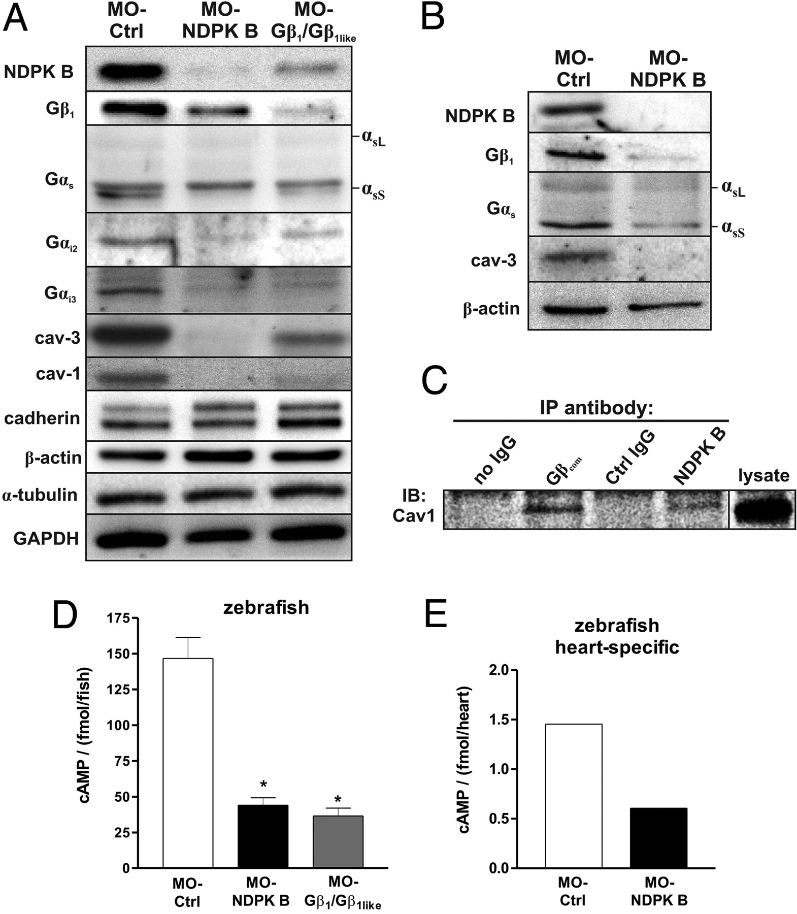
Fig. 5
Alterations of G protein content and cAMP formation in NDPK B and Gβ knockdown zebrafish embryos. (A and B) Representative immunoblots of zebrafish embryos at 72 hpf following knockdown of either NDPK B or Gβ1/Gβ1like in total fish lysates (A) and heart-specific extracts (B) using specific antibodies against the indicated G protein subunit and caveolin isoform. Positions of the long (αsL) and short (αsS) Gαs splice variants are indicated. Note that GαsS is migrating below a nonspecific immunoreactive band. Marker proteins (cadherin, β-actin, α-tubulin, GAPDH) representing different cell compartments are shown as loading and specificity control. (C) Co-immunoprecipitation of caveolin-1 with NDPK B and Gη. Endogenous Gβ or NDPK B was immunoprecipitated (IP) from zebrafish lysates (refer to Fig. 1C). Association of caveolin-1 was detected by immunoblotting (IB). A representative immunoblot is shown. (D) cAMP was measured in lysates from whole embryos at 72 hpf injected with the indicated MOs. Data are means ± SEM, n = 3, *, P < 0.05 vs. MO-Ctrl. (E) cAMP content of zebrafish hearts isolated from MO-Ctrl vs. MO-NDPK B-injected fishes with heart-specific GFP expression [Tg(cmlc2:gfp)] at 72 hpf. n = 365 hearts were pooled and lysed for each condition, respectively. The average of two independent experiments is shown.