
|
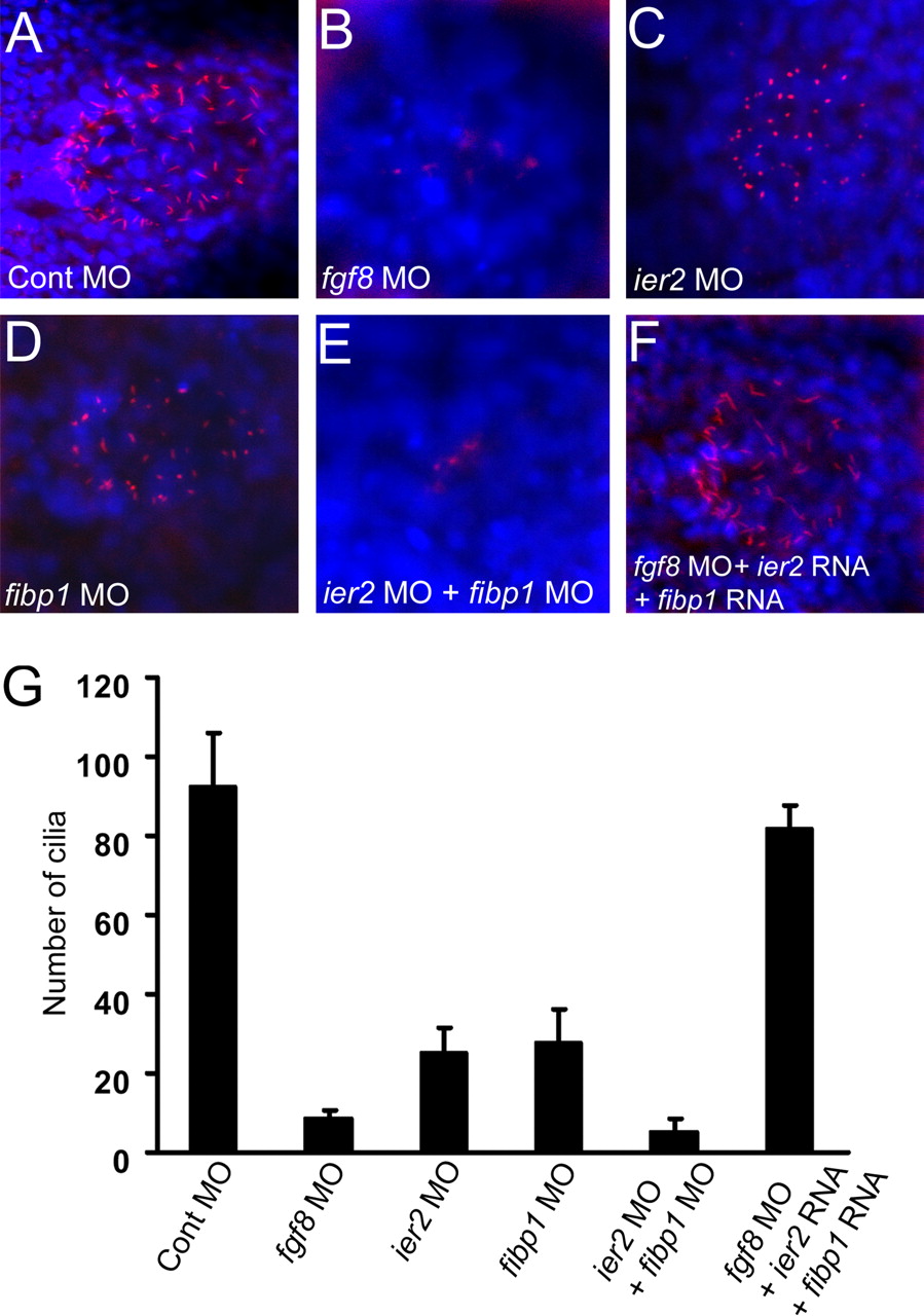
Fig. 5 Cilia formation in Kupffer′s vesicle depends on Ier2 and Fibp1. (A–F) Monocilia were detected with anti-acetylated tubulin antibody (red), and nuclei with DAPI (blue) in Kupffer′s vesicle at the 5-somite stage. fgf8 MO, 8 ng (B) and ier2 MO plus fibp1 MO (E) -injected embryos show dramatic reduction of cilia, compared with control (A). Moderate reduction of cilia by ier2 MO (C) or fibp1 MO (D) alone was observed. Loss of cilia was rescued by coinjection with ier2 RNA plus fibp1 RNA with fgf8 MO (F). For levels of MOs see legend to Fig. 4. (G) Number of cilia in Kupffer′s vesicle; 12 embryos were counted in each case corresponding to images in (A–F). n, notochord.