
|
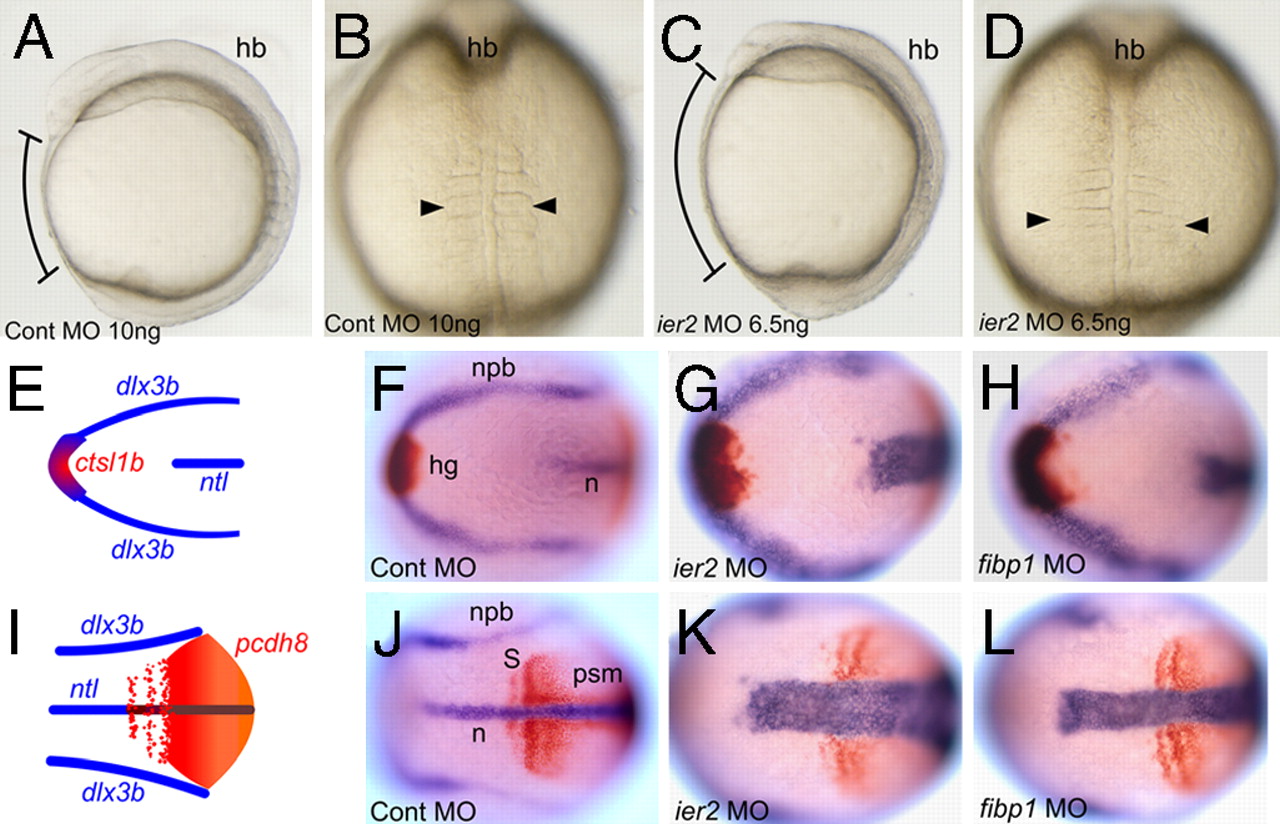
Fig. 2
Ier2 and Fibp1 are required for convergent extension. Control (A and B) and ier2 morphant (C and D). Lateral (A and C) and dorsal (B and D) views at the 5-somite stage. Brackets in A and C show the distance between head and tail, and arrowheads in B and D show the width of the somites. (E–L) Two-color in situ hybridization with marker genes at the 3-somite stage. Dorsal views of anterior (E–H) and posterior (I–L) region. (E and I) Drawings of a normal embryo indicating the patterns of the markers used, ctsl1b (red), dlx3b (blue), ntl (blue), and pcdh8 (red). (F and J) Control MO-injected embryo. (G and K) ier2 MO-injected embryo. (H and L) fibp1 MO-injected embryo. In the experimental embryos, notochord and somites are wider and extension along the A/P axis is reduced.