
Image
|
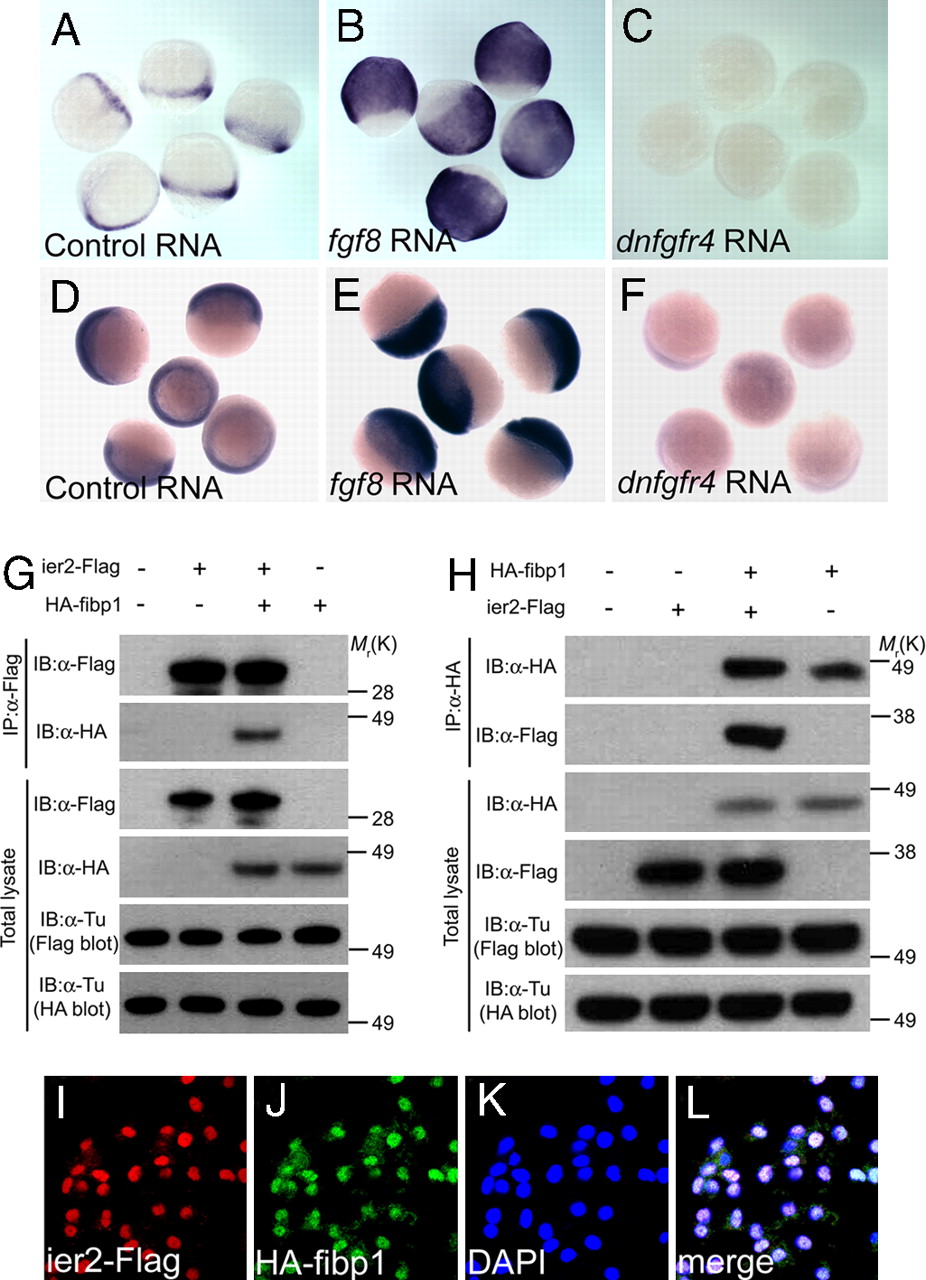
Figure Caption
Fig. 1
Ier2 and Fibp1 are Downstream of FGF. (A–F) Whole-mount in situ hybridization with ier2 (A–C) at 80% epiboly, and with fibp1 (D–F) at 60% epiboly stages after injection of control gfp RNA, 100 pg (A and D); fgf8 RNA, 5 pg (B and E); and dnfgfr4 RNA, 300 pg (C and F). (G and H) Physical interaction between Ier2 and Fibp1. Immunoprecipitation (IP) and immunoblotting (IB) are indicated. (I–L) Intracellular localization of Ier2 and Fibp1. Ier2 (I) and Fibp1 (J) are colocalized in the nucleus (K) after injection of epitope-tagged constructs into zebrafish embryos. (L) Merged image I–K
Acknowledgments
This image is the copyrighted work of the attributed author or publisher, and
ZFIN has permission only to display this image to its users.
Additional permissions should be obtained from the applicable author or publisher of the image.
Full text @ Proc. Natl. Acad. Sci. USA