
Image
|
Figure Caption
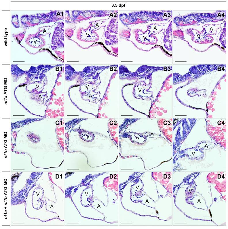
Fig. S5 Histological analysis of MO knockdown cardiac valves. Serial transverse histological sections through the atrioventricular valves of wild-type (A1–A4) or ≈2 ng morphant nf1a ATG (B1–B4), nf1b ATG (C1–C4), and nf1a + nf1b ATG (D1–D4) 3.5-dpf zebrafish embryos reveal no readily apparent defects at the resolution afforded to us by histological analysis (A, atrium; V, ventricle). (Scale bars: 50 μm.)
Acknowledgments
This image is the copyrighted work of the attributed author or publisher, and
ZFIN has permission only to display this image to its users.
Additional permissions should be obtained from the applicable author or publisher of the image.
Full text @ Proc. Natl. Acad. Sci. USA