
|
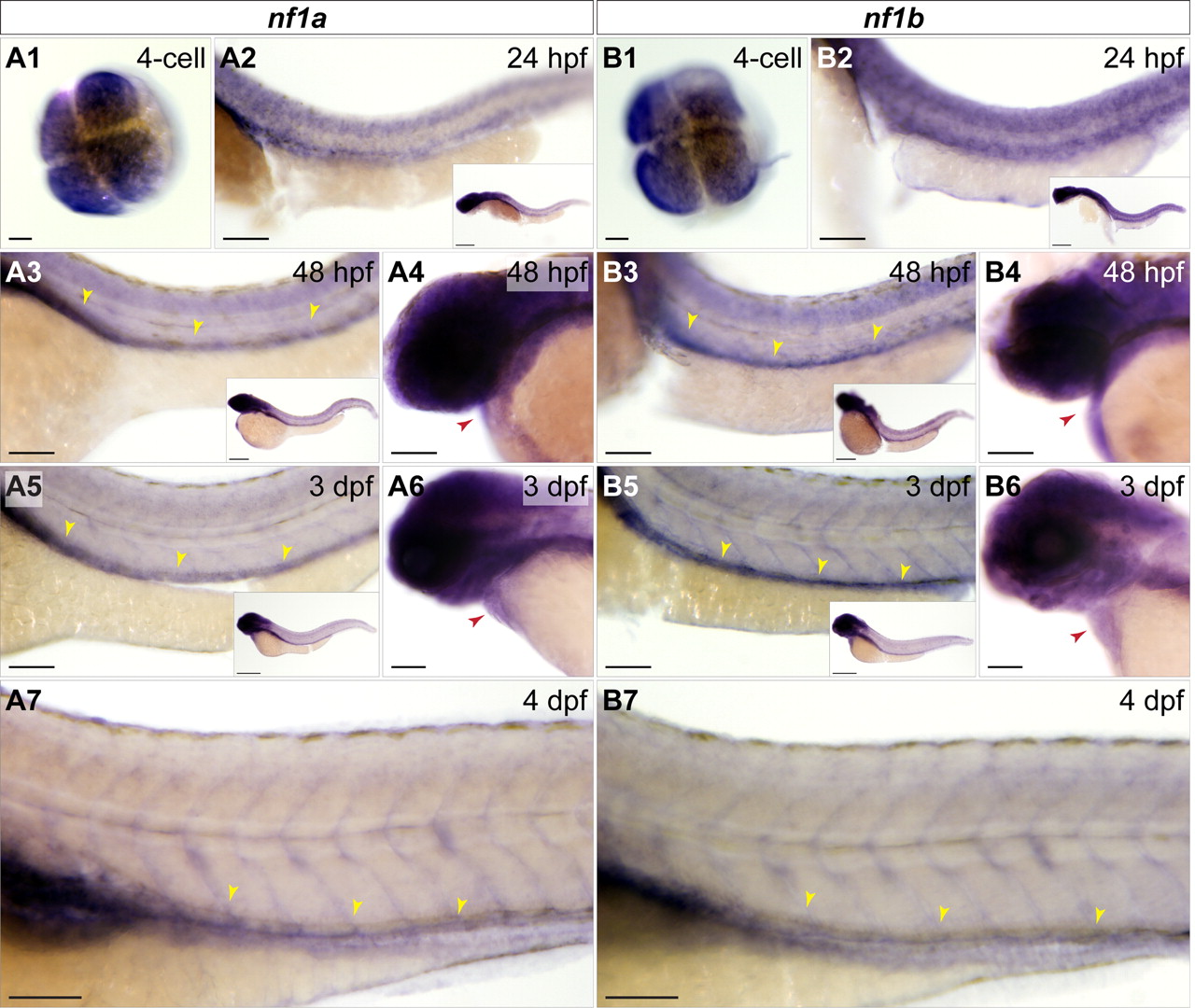
Fig. 2
nf1a and nf1b are expressed maternally and in the developing zebrafish cardiovascular system. Whole-mount in situ hybridization for nf1a and nf1b at the four-cell stage, 24 hpf, 48 hpf, 3 dpf, and 4 dpf. (A1 and B1) At the four-cell stage, nf1a and nf1b are expressed throughout the animal pole of the developing embryo. (A2 and B2) Both genes are expressed broadly at 24 hpf (Inset), with strong expression along the spinal cord. (A3 and B3) At 48 hpf, expression of nf1a and nf1b is noted in the head and regions of the anterior trunk (Inset). Spinal cord expression of both genes persists, and positive staining is observed along the dorsal vessel for nf1a and nf1b. (A4 and B4) Cardiac expression for both genes is observed at 48 hpf. (A5 and B5) Expression of nf1a and nf1b become progressively restricted to regions of the head at 3 dpf (Insets). nf1a and nf1b expression along the dorsal vessel (A5 and B5) and in the embryonic heart (A6 and B6) persist at 3 dpf. (A7 and B7) At 4 dpf, robust vascular staining is apparent for nf1a and nf1b. (Scale bars: 25 μm; 100 μm for insets.)