
Image
|
Figure Caption
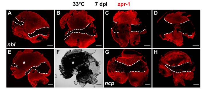
Fig. S5 Cone regeneration defect in nbl and ncp mutants at the restrictive temperature. (A–E, G, and H) Flat-mounted retinas at 7 dpl immunolabeled with zpr-1 (red). (A–E) One retina from each of 5 nbl mutants. (F) Bright-field image of E. (G and H) One retina from each of 2 ncp mutants. Dashed lines, light-damaged areas that have few or no zpr-1–labeled cones; we cannot determine from these preparations whether the rare scattered cones sometimes observed within the light-damaged areas survived the lesion or have regenerated. Asterisk, attached retinal pigment epithelium. (Scale bars: 300 μm.)
Acknowledgments
This image is the copyrighted work of the attributed author or publisher, and
ZFIN has permission only to display this image to its users.
Additional permissions should be obtained from the applicable author or publisher of the image.
Full text @ Proc. Natl. Acad. Sci. USA