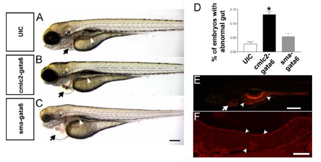
Fig. S6 Characterization of tissue-specific gata6 overexpression on development. (A–C) Pericardial edema (black arrow) results from injection of either SMA:Gata6 or CMLC2:Gata6, but only SMA:Gata6-injected embryos show an abnormal gut tube (white arrowhead) at 96 hpf. (D) The percentage of embryos exhibiting the abnormal gut morphology was analyzed by using Student’s t test (P < 0.05). A significantly higher percentage of embryos with abnormal gut development is found in SMA:Gata6 injected embryos. Bars,mean±SEM. *, P<0.05 (n>50 per group). (E) Lateral view of a control Tg (SMA: mCherry) transgenic embryo at 96 hpf. The heart (white arrow) and gut smooth muscle (white arrowheads) show expression of mCherry. (F) Longitudinal section of a 96 hpf Tg (SMA: mCherry) embryo shows that mCherry is specifically expressed in visceral smooth muscle cells (white arrowheads). (Scale bars, C: 200 μm; E: 400 μm; F: 50 μm.)
Image
Figure Caption
Acknowledgments
This image is the copyrighted work of the attributed author or publisher, and
ZFIN has permission only to display this image to its users.
Additional permissions should be obtained from the applicable author or publisher of the image.
Full text @ Proc. Natl. Acad. Sci. USA

