
|
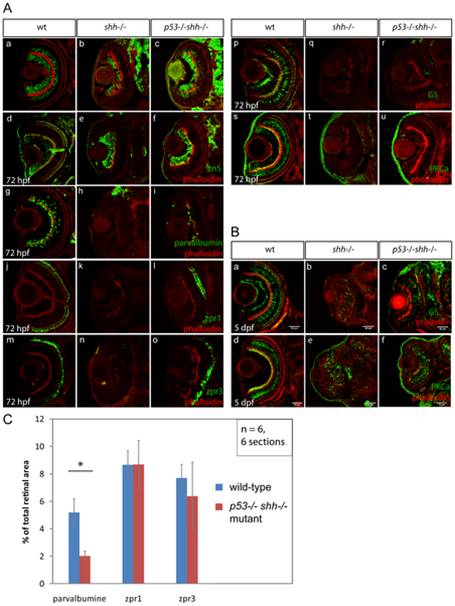
Fig. 9 Differentiation of retinal cell types was assessed using specific antibodies (A, B). The sections are oriented with their anterior side to the top. All sections were stained with phalloidin-Alexa568 (A, a–u; B, a–f) to visualize retinal lamination. Staining for HuC neuronal antigen labelled ganglion and amacrine cells in the wild-type retina (A, a), ganglion and very few amacrine cells in the shh-/- mutant retina (A, b). In the p53-/-shh-/- retina, however, labelling of both ganglion cells and amacrine cells was partially restored (A, c). Ganglion cells labelled by an anti-zn5 antibody were present in comparable numbers in wild-type (A, d), shh-/- mutant (A, e) and p53-/-shh-/- mutant retinas (A, f). Amacrine cells labelled by anti-parvalbumine antibody were present in the wild-type retina (A, g), absent in the shh-/- mutant (A, h) and partially rescued in the p53-/-shh-/- mutant retina (A, i). Red-green double cones (zpr1 antibody labelling) and rod photoreceptors (zpr3 antibody labeling) were present in the wild-type retina (A, j, m), absent in the shh-/- mutant retina (A, k, n) and rescued in the p53-/-shh-/- mutant retina (A, l, o). Staining for glutamine synthetase (GS) labelled Müller glia cells in wild-type embryos (A, p). In both shh-/- and p53-/-shh-/- retinas, very few Müller glia were present (A, q, r). Bipolar cells were labelled in the wild-type retina by anti-PKCα antibody staining (A, s), but absent in shh-/- and p53-/-shh-/- retinas (A, t, u). At 5 dpf, Müller glia and bipolar cells were detected in shh-/- and p53-/-shh-/- retinas (B, b,c,e,f) but in lower numbers than in the wild-type retina (B, a,d). All stainings were performed on at least 6 embryos for each genotype and representative images are shown. (C) Quantitation of rescue of amacrine cells (parvalbumine labelling), red-green double cone (zpr1) and rod photoreceptors (zpr3) in the p53-/-shh-/- mutant retina by plotting ratios of segmented area for each staining to the total retina area. Amacrine cells occupy a significantly larger relative area in the wild-type than in the p53-/-shh-/- mutant retina (* on top of the bars, t-test, P-value<0,05), whereas relative areas of both types of photoreceptors are not significantly different in wild-type and p53-/-shh-/- mutant retinas.