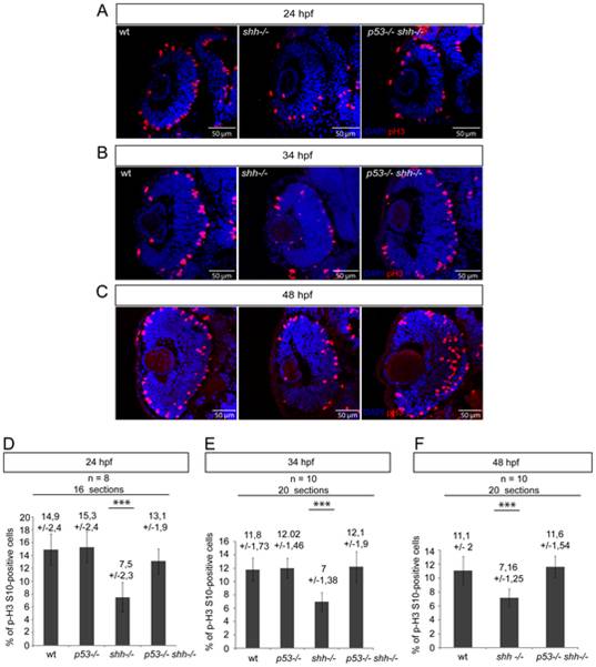
Fig. 7 pH 3/DAPI staining of wild-type, shh-/- and p53-/-shh-/- retinas was done at 24 hpf (A), 34 hpf (B) and 48 hpf (C). At all stages, the mitotic index in the shh-/- retina was lower than in the wild-type retina, while in the p53-/-shh-/- retina, the mitotic index was comparable to that of the wild-type retina. (D) Statistical analysis of mitotic indices in wild-type, p53-/-, shh-/- and p53-/-shh-/- retinas at 24 hpf, the indices being comparable in wild-type (14,9%, SD = 2,4%), p53-/- (15,3%, SD = 2,4%), and p53-/-shh-/- (13,1%, SD = 1,9%) embryos and significantly higher than in the shh-/- mutant retina (7,5%, SD = 2,3%). (E) Statistical analysis of mitotic indices in the wild-type, p53-/-, shh-/- and p53-/-shh-/- retinas at 34 hpf, comparable in wild-type (11,8%, SD = 1,73%), p53-/- (12,02%, SD = 1,46%), and p53-/-shh-/- (12,1%, SD = 1,9%) retinas, whereas the mitotic index in the shh-/- mutant retina was almost twice lower (7%, SD = 1,38%). (F) Statistical analysis of mitotic indices in wild-type, shh-/- and p53-/-shh-/- retinas at 48 hpf, the indices being comparable in wild-type (11,1%, SD = 2%) and p53-/-shh-/- (11,6%, SD = 1,54%) embryos and significantly higher than in the shh-/- mutant retina (7,16%, SD = 1,25%). Either 8 (D) or 10 (E, F) embryos (2 retinal sections per embryo) were analysed for each genotype and mean +/- standard deviation indicated above the bars of mitotic index statistics. Asterisks (***) on top of shh-/- mutant bars indicate their significant statistical differences from other samples (t-test, P-value<0,001).
Image
Figure Caption
Acknowledgments
This image is the copyrighted work of the attributed author or publisher, and
ZFIN has permission only to display this image to its users.
Additional permissions should be obtained from the applicable author or publisher of the image.
Full text @ PLoS One

