
|
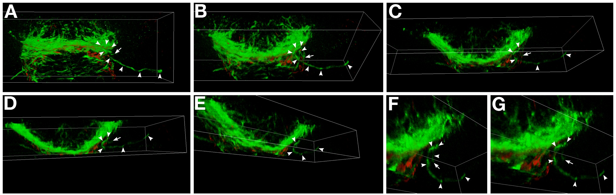
Fig. S1 Three-dimensional analysis of optic nerve defects in ppp1r12a mutants. A-G: Selected maximum intensity projections that correspond to the 3-D rotation of the postoptic commissure and developing optic nerves. In ppp1r12a mutants at this time (40 hr) the optic nerves are seen at or near the midline, but one of the optic nerves turns away from the midline and grows ipsilateral (axon, arrowheads; leading edge, arrow). B-D: This retinal ganglion axon pathfinding error is more obvious when the forebrain is rotated downward along the X axis. E-G: Rotating along the Y axis. F,G: Higher magnification of the ipsilaterally growing axon. Axons, green (αAcetylated Tubulin); astroglia, red (anti-Gfap). This data corresponds to the movie presented in Supplemental Movie 1