Image
Figure Caption
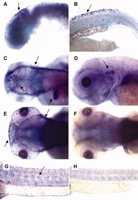
Fig. 5 In situ hybridization of Cx30.3 mRNA in embryos. Embryos were stained by whole-mount in situ hybridization with a cx30.3 antisense riboprobe. A,B: Signal was detected (and highlighted with arrows) in 1 days postfertilization (dpf) stage embryos, in the otic vesicle (A) and dorsal fins (B). C-H: At 2 dpf (C,G,H) or 3 dpf (D-F), cx30.3 mRNA signal was readily detected in the skin (C), the inner ear (D), the lateral line (E,G), and the fins (C). F,H: Sense-probe controls are shown.
Figure Data
Acknowledgments
This image is the copyrighted work of the attributed author or publisher, and
ZFIN has permission only to display this image to its users.
Additional permissions should be obtained from the applicable author or publisher of the image.
Full text @ Dev. Dyn.

