
|
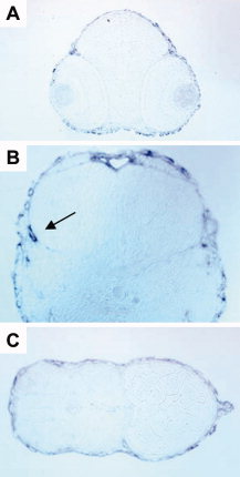
Fig. 6 Sections of cx30.3 in situ hybridization stained embryos. Embryos were stained by whole-mount in situ hybridization with cx30.3 antisense riboprobe, embedded in plastic and 5-μm sections were cut with a microtome. Only sections from embryos at 2 days postfertilization (dpf) are shown, the staining pattern in later stage embryos is similar. A: Transverse cross-section of embryonic eyes, showing a lack of staining of the probe. B: The developing inner ear with probe labeling the otic vesicle (arrow). C: A transverse section of the embryonic fish tail and fin, with probe accumulating around the peripheral skin and on the fin (far right).