
|
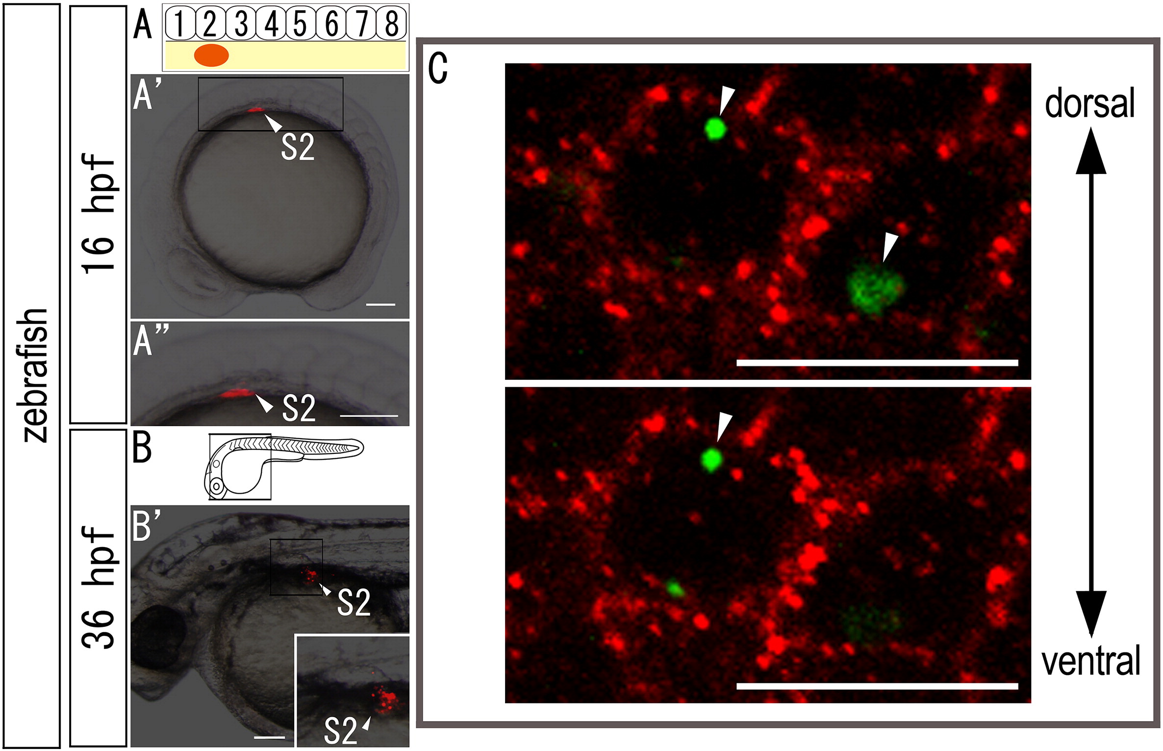
Fig. S2 Cell lineage analysis in zebrafish embryos. (A-B) Locations of DiI-labeled cells (A) are indicated by arrowheads. (A) Schematic diagrams showing the locations at which DiI (red) was applied. (A′) DiI was applied to the lateral plate mesoderm cells opposite somite 2 (A′) in 16 hpf zebrafish embryos. (A3, B′) Higher magnification of the labeled regions shown in (A′) and (B), respectively. (C) Intracellular localization of aggregated Qdot granules in the cytoplasm. Confocal micrographs were obtained through the mesodermal cells labeled with Qtracker. Embryos were stained with anti-β-catenin antibody, a marker of the membrane (Nagafuchi, A., 2001. Molecular architecture of adherens junctions. Curr Opin Cell Biol. 13, 600-3). Note that the aggregated Qdot nanocrystals are delivered to the intracellular domains (arrowheads) of mesodermal cells. Scale bars: 100 μm for panels (A) and (B); 5 μm for a panel (C).
Reprinted from Developmental Biology, 347(1), Murata, Y., Tamura, M., Aita, Y., Fujimura, K., Murakami, Y., Okabe, M., Okada, N., and Tanaka, M., Allometric growth of the trunk leads to the rostral shift of the pelvic fin in teleost fishes, 236-245, Copyright (2010) with permission from Elsevier. Full text @ Dev. Biol.