
|
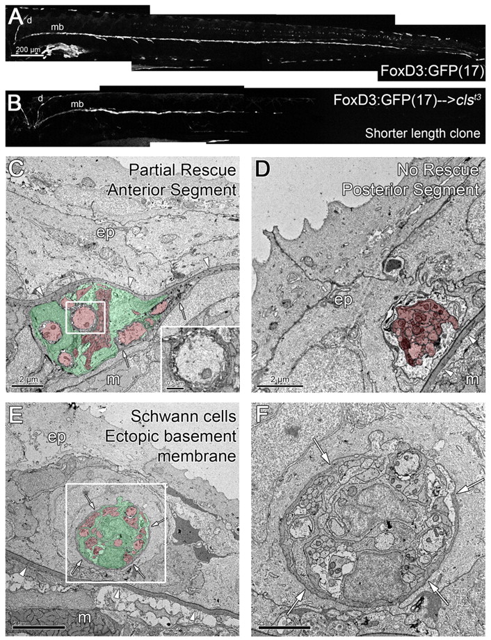
Fig. 5 Shorter length clones of transplanted wild-type Schwann cells can partially rescue the position of sox10/clst3 mutant nerves. (A) Wild-type 5 dpf embryo carrying the FoxD3:GFP(17) transgene, labeling the PLLn [dorsal (d) and midbody (mb, tracts], motor nerves and some pigment cells. (B) A clone of wild-type, FoxD3:GFP(17) cells that are present on the dorsal and anterior regions of the midbody tract of the PLLn. (C,D) Micrographs of the anterior (C) and posterior (D) regions of a partial length clone of rescued Schwann cells, as shown in B. (C) Wild-type Schwann cells rescue the position of the mutant nerve, but a part of the old basement membrane remains (arrows). Inset in C shows the presence of myelinated axons. (D) The posterior region of the nerve lacks Schwann cells and is not rescued, remaining within the epidermis. (E,F) In this chimera, wild-type Schwann cells failed to rescue the position of the PLLn, but induced the formation of an ectopic basement membrane surrounding the nerve in the epidermis (arrows). (F) Higher magnification of boxed area in E. Arrowheads indicate the epidermal basement membrane; arrows indicate ectopic basement membrane. Scale bars: 200 μm for A,B; 2 μm for C,D,F; 5 μm for E. Schwann cells are pseudocolored green and axons red in C-E. Abbreviations: d, dorsal tract; mb, midbody tract; ep, epidermis; m, muscle.