
Fig. 5
- ID
- ZDB-IMAGE-101104-30
- Publication
- Liu et al., 2010 - A dual role for ErbB2 signaling in cardiac trabeculation
- All Figures
- Figures for Liu et al., 2010
|
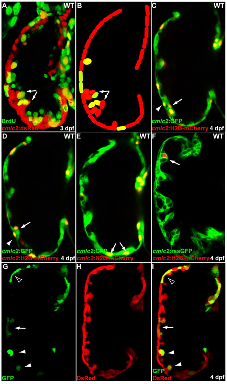
Fig. 5 Initiation of cardiac trabeculation appears to involve directional cardiomyocyte migration (delamination) rather than oriented cell division. (A,B) Tg(cmlc2:dsRed) embryos at 60 hpf were exposed to BrdU for 24 hours. BrdU labeling shows that not all trabecular cells (arrows) are BrdU-positive. (C-F) cmlc2:H2B-mCherry reporter construct was injected into one-cell-stage Tg(cmlc2:GFP) (C-E) or Tg(cmlc2:rasGFP) (F) embryos and the distribution of mCherry-positive cardiomyocytes was monitored at 4 dpf. (C,D) Two optical sections of the same heart show that the lineage marker mCherry labels compact layer cardiomyocytes (arrowheads) as well as some of their immediate trabecular neighbors (arrows). (E,F) Trabecular cardiomyocytes (arrows), but not their compact layer neighbors, are labeled in another heart. (G-I) Genetic clones generated by crossing Tg(cmlc2:CreER) fish with Tg(bactin2:loxP-DsRed-STOP-loxP-GFP) reporter animals were found to contain both compact and trabecular cardiomyocytes (arrow), only compact cardiomyocytes (empty arrowhead) or only trabecular cardiomyocytes (arrowheads).