
|
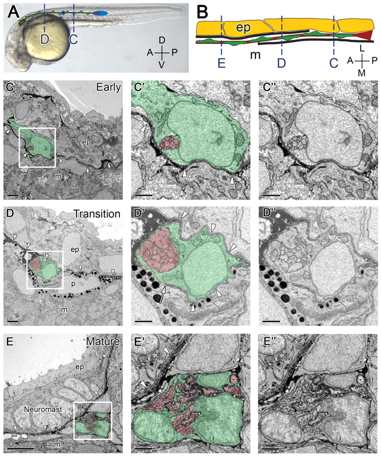
Fig. 1 The PLLn invades the epidermal basement membrane. (A) 28 hpf embryo with schematic of the PLLn superimposed. Schwann cells are green, axons and the ganglion are red, and the primordium and a pre-neuromast (asterisk) are blue. Broken lines refer to approximate location of cross-sections shown in C and D. (B) Schematic representation of the transition of the PLLn across the epidermal basement membrane. Basement membrane is shown in black, Schwann cells in green, axons in red, epidermal cells in yellow; only one of two epidermal cell layers is shown. Broken lines correspond to cross-sections in C-E. (C) At 28 hpf, the distal segments of axons and associated Schwann cells of the PLLn are located within the epidermis (n=6). Arrowheads indicate the epidermal basement membrane. (D) At locations more anterior to that shown in C at 28 hpf, the PLLn is found in a transition state, with basement membrane located on both sides of the nerve (arrowheads). Arrow indicates the end of the basement membrane (n=6). (E) By 3 dpf, the PLLn is found at its mature location, medial to the epidermal basement membrane (arrowheads, n=9). A neuromast is present within the epidermis. (C′,C′,D′,D′,E′,E′) Higher magnification of boxed regions in C,D,E, with and without pseudocoloring. Scale bars: 2 μm in C,D; 5 μm in E; 1 μm in C′,D′,E′. Schwann cells are pseudocolored green, axons are red. Abbreviations: D, dorsal; V, ventral; A, anterior; P, posterior; L, lateral; M, medial; ep, epidermis; m, muscle; p, pigment.