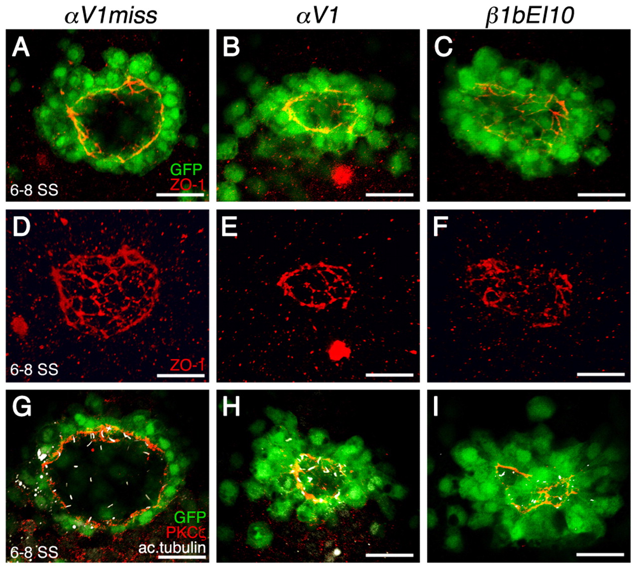
Fig. 8 KV lumen does not properly form in αV and β1b morphants. Confocal images of Tg(sox17:GFP)-expressing embryos (green) are shown. (A-C,G-I) Single focal planes at the center of the DFC cluster immunolabeled with anti-ZO-1 antibody (red; A-C) or anti-aPKC-ζ antibody (red) and anti-acetylated tubulin antibody (white; G-I). (D-F) 3D renderings of anti-ZO-1 labeled embryos of A-C. Dorsal views of 6-8 SS embryos are shown in all panels, anterior to the top. Embryos injected with αV1miss control MO developed a large fluid-filled lumen (A,G) that had a uniform ZO-1-labeled tight junction lattice within the DFC-derived lining of the KV (D). However, in αV (B,H) and β1b (C,I) morphants, DFCs did not aggregate properly, yielding a dysmorphic ZO-1 lattice (E,F). Anti-aPKCζ staining shows that KV cells in αV1miss morphants were polarized (G), but not in αV (H) and β1b morphants (I). Scale bars: 30 μm.
Image
Figure Caption
Figure Data
Acknowledgments
This image is the copyrighted work of the attributed author or publisher, and
ZFIN has permission only to display this image to its users.
Additional permissions should be obtained from the applicable author or publisher of the image.
Full text @ Development

