
|
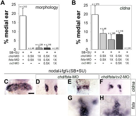
Fig. 6 Development of medial otic tissue requires mesendodermal Bmp-antagonists. A, B: Frequency of inhibitor-treated embryos producing medial otic vesicles as determined by morphological criteria (A) or expression of cldna (B). Embryos obtained from outcrosses of oep+/- heterozygotes were treated with 70 μM SB431542 + 1 μM SU5402. Where indicated, embryos were also injected with 2.5 ng chd-MO, 5 ng fsta-MO, and 5 ng of cv2-MO2 (1X), or with half-doses of morpholino (0.5X). Data are presented as the mean and SEM of 2–8 independent experiments, with total sample sizes indicated. C–F: Representative specimens showing cldna expression at 24 h (C–F). G, H: Expression of fsta at 11 hpf shows that paraxial cephalic mesendoderm is not ablated by the combination of SB431542, SU5402, and 1X morpholinos. All images are dorsal views (anterior to top). Scale bar = 50 μm.