
|
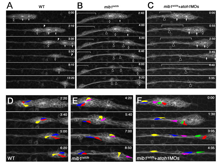
Fig. S4 Knockdown of atoh1 does not restore the stability of deposited neuromasts in mib1 mutants. Time-lapse imaging of pLLp migration and proneuromast deposition starting at <28 hpf. In D-F, to track the movement of individual cells, three or four cells are labeled with different colors. (A) In wild-type embryos, a maturing neuromast at the trailing end of the pLLp (black arrows) slowed down and separated from the remaining pLLp following a break (white arrowheads) in the column of cells in which maturing neuromasts (white arrows) continue to migrate. See Movie 1. (B) In mib1ta52b mutants, initiation of rosette formation is not significantly affected; however, maturing neuromasts do not form stable epithelial rosettes. Black arrowheads show cells dissociating and reassociating with others after they break away from a depositing neuromast (black arrows). The white brackets show spread out cells from a previously deposited neuromast progressively trying to reassociate to form a more cohesive structure. See Movie 2. (C) Whereas knockdown of atoh1a and atoh1b function in mib1ta52b mutants significantly improved overall cohesion and migration of pLLp, it reduced cohesive interactions between cells that disassociate from deposited neuromasts. Disassociated cells within white brackets failed to reassociate and eventually became indistinguishable from interneuromast cells. See Movie 5. (D) In the wild-type, the labeled cells were maintained in the epithelial rosette during proneuromast deposition. (E) In mib1ta52b mutants, a subset of cells disassociated from a neuromast (red, blue) and eventually reassociated with cells from a previous neuromast, while others (yellow) remained in the original cell cluster. The pink cell was released from the original rosette reassociated with the pLLp. As a result, the size of the epithelial rosette at the most trailing end of the pLLp became smaller. (F) In mib1ta52b mutants injected with atoh1MOs, there was an important change in the behavior of the cells in maturing neuromasts. Although individual cells continued to disassociate from maturing neuromasts (blue, yellow, pink and green cells), there was no tendency to try and reassociate and form new clusters with previously disassociated cells or previously deposited neuromasts. Instead, they remained as individuals and became indistinguishable from other interneuromast cells normally seen between deposited neuromasts.