
|
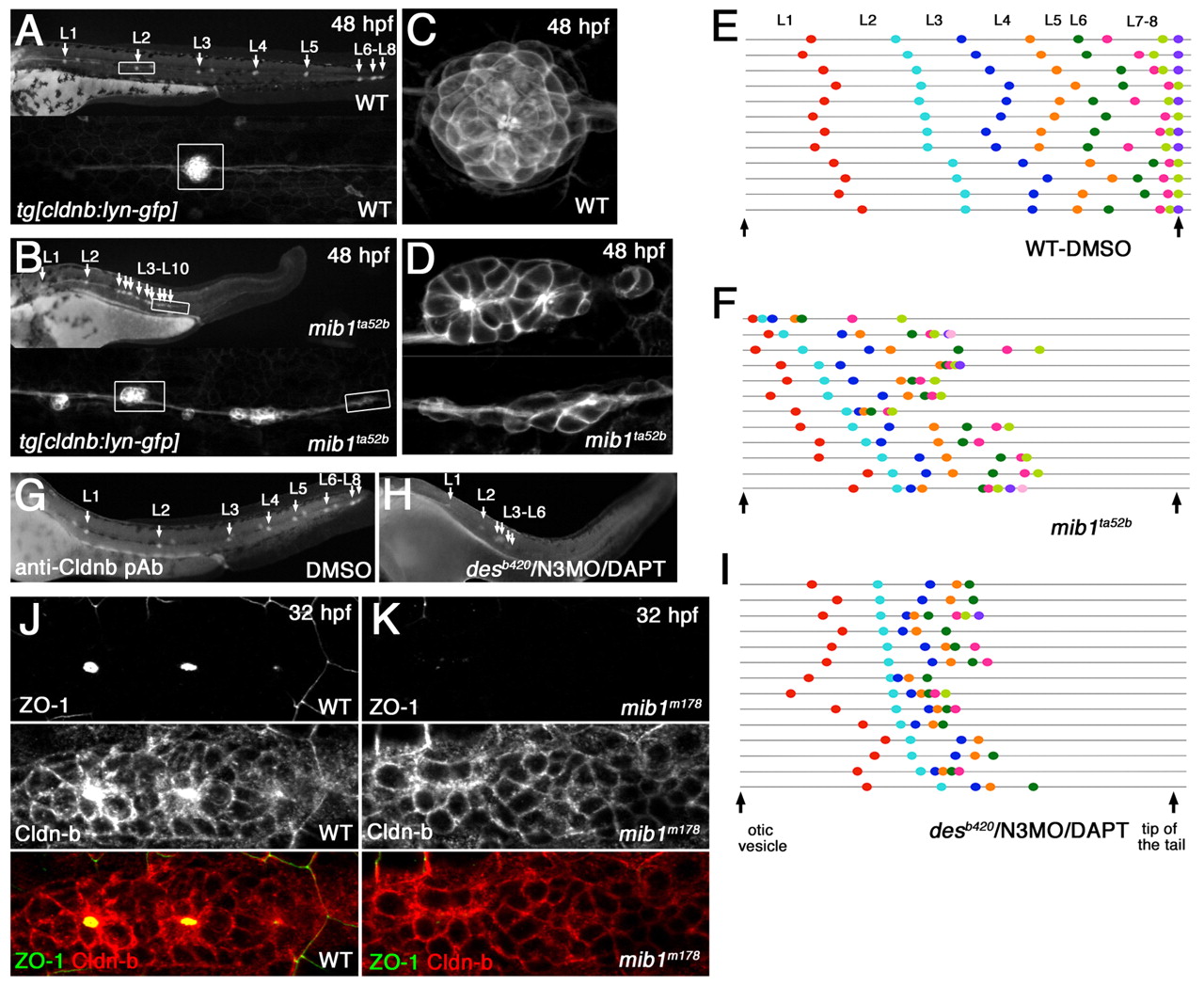
Fig. 1 Loss of Notch signaling causes aberrant neuromast deposition and posterior lateral line primordium (pLLp) migration. (A-D) The distribution and morphology of deposited neuromasts/cell clusters (L1 to L10) in (A,C) wild-type (WT) and (B,D) mib1ta52b tg[cldnb:lynGFP] zebrafish embryos at 48 hpf. (C,D) Epithelial rosettes are disorganized in mib1ta52b mutants. (E,F,I) The pattern of neuromast/cell cluster deposition at 48 hpf in individual wild-type (E), mib1ta52b (F) and desb420/N3MO/DAPT (I) embryos. Colored dots show the position of each deposited neuromast/cell cluster relative to the otic vesicle and tail tip (vertical arrows). Individual embryos are represented on separate lines in an order determined by the relative position of L2. (G,H) The distribution of deposited neuromasts/cell clusters in DMSO-treated (G) and desb420/N3MO/DAPT (H) embryos at 48 hpf. (J,K) There is no central accumulation of ZO1 and Cldnb accumulation is delayed in mib1m178 neuromasts (K) when compared with the wild type (J) at 32 hpf.