
Image
|
Figure Caption
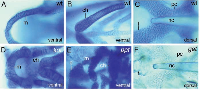
Fig. 8 Photomicrographs of cartilaginous elements of wild-type (A-C), knorrig (D), pipe tail (E) and geist (F) mutant larvae. (A,D,E) show ventral close-up views of Meckel’s and the ceratohyal. In knorrig mutant larvae (D), Meckel’s is smaller but thicker and chondrocytes grow along the edges. In pipe tail mutant embryos, Meckel’s lies further to the posterior and all elements consist of smaller but more numerous cells. The ceratohyal is relatively bigger than the other elements. Dorsal views of the neurocranium (C,F) indicate that chondrocytes of geist mutant larvae hardly stain with Alcian blue.
Figure Data
Acknowledgments
This image is the copyrighted work of the attributed author or publisher, and
ZFIN has permission only to display this image to its users.
Additional permissions should be obtained from the applicable author or publisher of the image.
Full text @ Development