
|
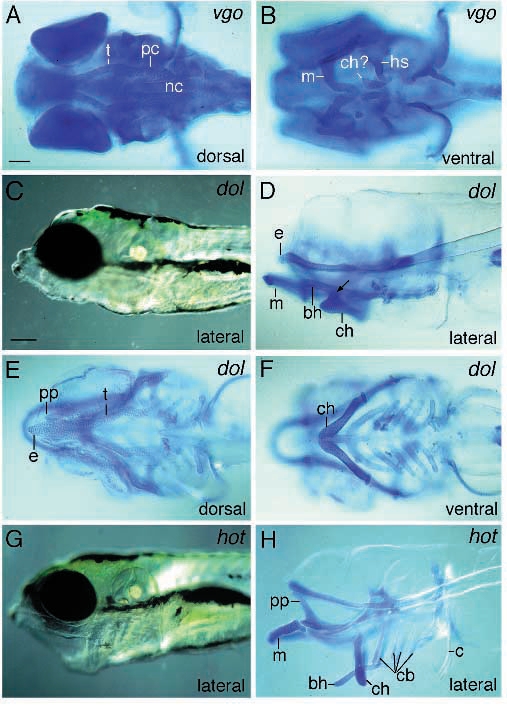
Fig. 5 Photomicrographs of van gogh (tm208) (A,B), dolphin (C-F) and hanging out (G,H) mutant larvae. In van gogh mutant embryos all elements are severely malformed. A ventral view (A) reveals that Meckel’s is inverted and the ceratohyals are not clearly identifiable any more. The posterior arches are reduced or absent. The ear capsules are misshapen, as can be seen from a dorsal view (A). Live dolphin mutant larvae are characterized by a lack of tissue dorsal to the ethmoid plate (C). The ethmoid plate is arrow-like in shape and consists of fewer chondrocytes anteriorly (E). Lateral (D) and ventral (F) views of stained larvae show that the the ceratohyals fuse dorsally with each other and the overlying basihyal. In larvae homozygous for hanging out, the mouth always stays open (G) and the pharyngeal skeleton points ventrally (H). Scale bars: 200 μm (C and G) and 100 μm (A,B,D,E,F and H).