
|
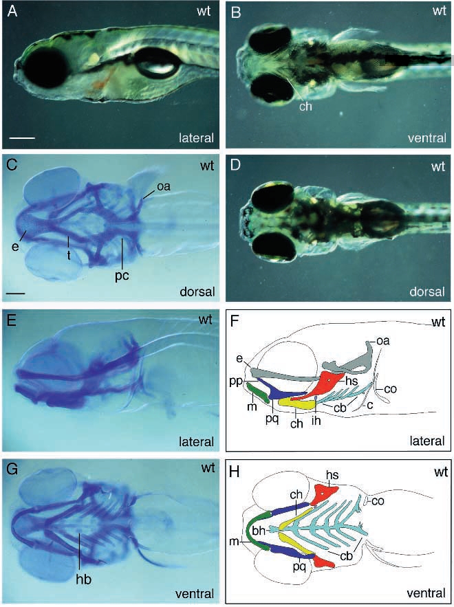
Fig. 1 Photomicrographs and schematic drawings of live (A,B and D) and Alcian blue-stained (C,E,G) wild-type larvae. Color-coded digrams (F,H) of the stained larvae shown in E and G depict distinct cartilaginous elements of the head and pectoral girdle. In H the neurocranium is omitted. C and D present dorsal views, whereas B and G show ventral views of live and stained larvae, respectively. A,E and F show lateral views of these larvae. Abbreviations used throughout all figures: bb, basibranchial; bh, basihyal; ch, ceratohyal; co, coracoid of pectoral girdle; cb, ceratobranchial; c, cleithrum; e, ethmoid plate; ff, facial foramen; hb, hypobranchial; hs, hyosymplectic; m, Meckel’s cartilage; oa, occipital arch; op, opercle; pc, parachordal; pp, pterygoid process of the palatoquadrate; pq, palatoquadrate; t, trabeculae; te, teeth. Color code used in all diagrams: green, Meckel’s; blue, platoquadrate; red, hyosymplectic; yellow, ceratohyal; light blue, interhyal; light green, arches 3-5; grey, neurocranium or ectopic cartilages; white, coracoid of pectoral girdle. Scale bars: A,B,D, 200 μm; C,E-H: 100 μm.