
|
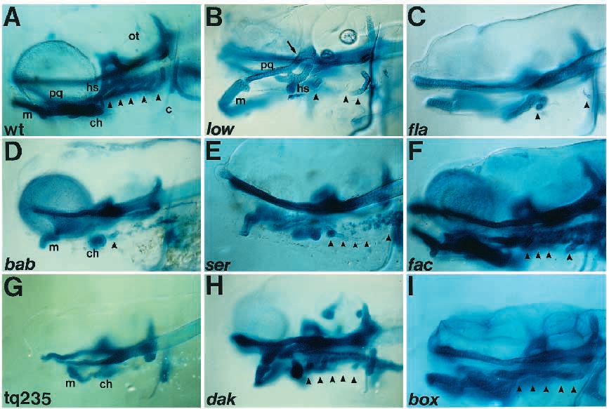
Fig. 3 Skeletal defects in mutants (lateral view). Cartilage in 6-day-old larvae is stained with Alcian blue and whole-mounted. Arrowheads indicate the positions of branchial arches. (A) Wild type, wt. There are five branchial arches, P3-P7. (B) low. Two branchial arches, P4 and P5, are absent. Meckel’s cartilage hangs ventrally, the hyosymplectic is reduced and the palatoquadrate makes ectopic contact with the neurocranium (arrow). (C) fla. Branchial arches P4-P6 are absent. (D) bab. All branchial arches are absent except the basibranchial of P3. Meckel’s cartilage hangs ventrally and the ceratohyal is reduced. The neurocranium is severely reduced. (E) ser. One branchial arch, P6, is absent. The ceratobranchial of P3 and basibranchials of P4 and P5 are present, as is a single tooth of P7. (F) fac. P6 is absent. (G) tq235. All branchial arches are absent. Anterior arches and the neurocranium are severely reduced. (H) dak. All branchial arches are reduced. Meckel’s cartilage hangs ventrally. (I) box. All branchial arches are reduced.