
Fig. 2
|
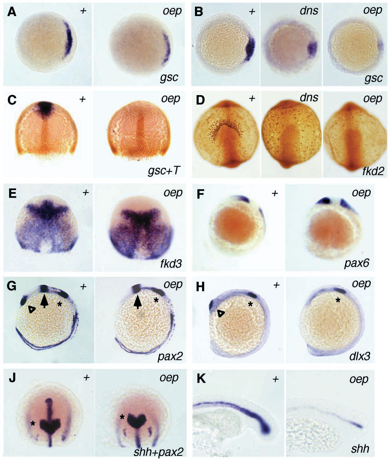
Fig. 2 Altered gene expression in mesoderm and neuroectoderm of oep and dns mutants revealed by in situ hybridization (A-C, E-K) and immunostaining (C,D). In each section, wild type (+) is to the left, mutants to the right. (A) gsc, 40% epiboly, animal view, dorsal right. (B) gsc, shield, animal view, dorsal right. (C) gsc mRNA in blue and Ntl protein (T) in brown, 70% epiboly, dorsal view. (D) fkd2, 10-somite stage, view on head: fkd2-positive presumptive hatching gland cells are absent in oep mutants. In dns mutants, very small fkd2-positive cells can be found at the anterior border of the hypoblast rather than in adjacent regions of the yolk sac. (E) fkd3, 70% epiboly, dorsal view: fkd3 expression is missing anterior to the diencephalic-mesencephalic boundary in oep mutants. (F) pax6, 3-somite stage, lateral view: the pax6 expression domain in the presumptive diencephalon reaches the anterior border of the CNS in oep mutants. (G) pax2, 12-somite stage, lateral view: pax2 expression is normal in the otic placode (asterisk) and in the midbrain (arrow) while the expression domain in the optic stalk (triangle) is missing. (H) dlx3, 12-somite stage, lateral view: the dlx3 expression is normal in the otic placode (asterisk) but absent in the olfactory placode (triangle) in oep mutants. (J) shh and pax2, tailbud-stage, view on head region: no shh expression can be detected in the brain anterior of the pax2-positive midbrain (asterisk). The axial transcripts posterior of the midbrain are located in the notochord. (K) shh, 22 hours, lateral view on tail region: shh expression is normal in the slightly undulated notochord but absent in the ventral cells of the spinal cord.